

doi: 10.1186/s40478-022-01419-3.
Disseminated diffuse midline gliomas, H3K27-altered mimicking diffuse leptomeningeal glioneuronal tumors: a diagnostical challenge!
Affiliations
- PMID: 35986414
- PMCID: PMC9392342
- DOI: 10.1186/s40478-022-01419-3
Item in Clipboard
Disseminated diffuse midline gliomas, H3K27-altered mimicking diffuse leptomeningeal glioneuronal tumors: a diagnostical challenge!
Acta Neuropathol Commun.
.
No abstract available
Conflict of interest statement
The authors declare that they have no conflict of interest directly related to the topic of this article.
Figures
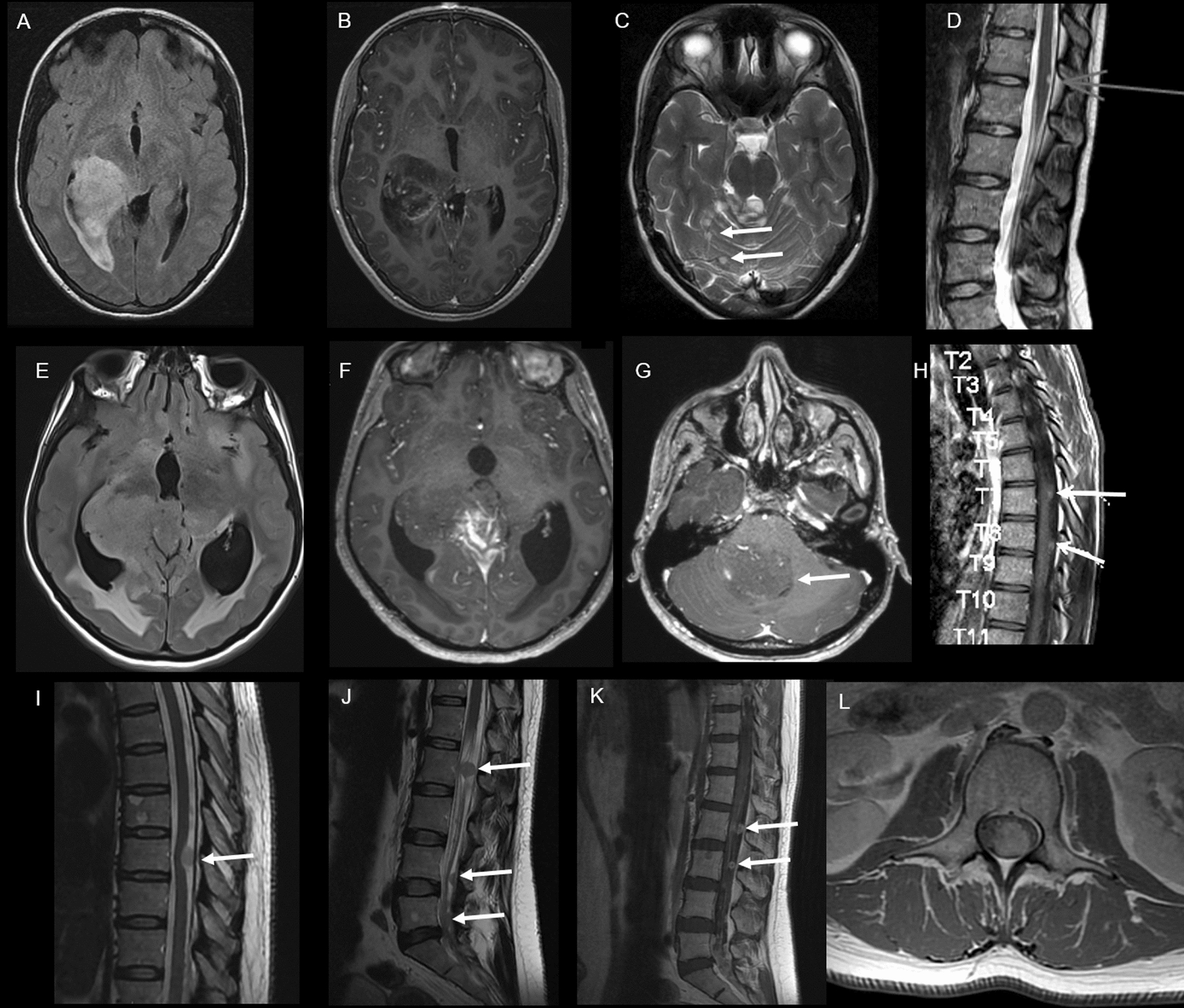
Radiological features. Case #1 A Axial FLAIR brain MRI shows a hyperintense infiltrative lesion of the right thalamus extended to the right lateral ventricle. B Axial contrast-enhanced T1-weighted brain MRI shows a heterogeneous enhancement after gadolinium injection. C Axial T2-weighted brain MRI shows other nodular FLAIR hyperintensities of the cerebellum (arrows). D Sagittal T2-weighted spine MRI shows a hyperintense peripheral lesion of the spinal cord (arrow). Case #2 E Axial FLAIR-weighted brain MRI shows a hyperintense lesion of the right thalamus extended to the third ventricle and the right hippocampus. F Axial contrast-enhanced T1-weighted brain MRI shows a heterogeneous enhancement of this lesion. G Axial contrast-enhanced T1-weighted brain MRI shows an intraventricular localization in the fourth ventricle (arrow). H Sagittal contrast-enhanced T1-weighted spine MRI shows multiple spinal leptomeningeal lesions. Case #3 I Sagittal T2-weighted spine MRI shows a thoracic hyperintense leptomeningeal lesion. J Sagittal T2-weighted lumbar MRI shows multiple lumbar intradural lesions, attached to nerve roots and in the lower end of the dural sac. K Sagittal and L Axial contrast-enhanced T1-weighted lumbar MRI show an enhancement of these lesions. FLAIR: Fluid Attenuated Inversion Recovery
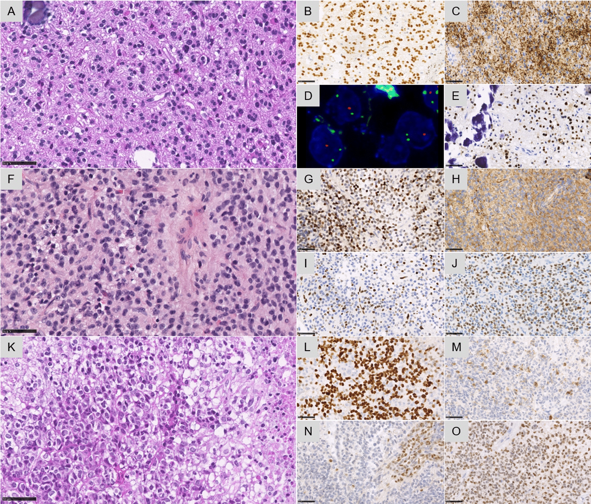
Histopathological and molecular features. Case #1 A A glial proliferation with oligo-like features and one microcalcification (HPS, magnification × 400). B Diffuse expression of Olig2 (magnification × 400). C Diffuse synaptophysin immunoreactivity without true neuropil islands (magnification × 400). D 1p deletion by FISH analysis (green signal for 1q25 and orange signal for 1p36, magnification × 400). E EZHIP overexpression in all tumor cells (magnification × 400). Case #2 F A glial proliferation with astrocytic features (magnification × 400). G Diffuse expression of Olig2 (magnification × 400). H Diffuse synaptophysin immunoreactivity without true neuropil islands (magnification × 400). I Loss of the trimethylation H3K27me3 in tumor cells (magnification × 400). J EZHIP overexpression in all tumor cells (magnification × 400). Case #3 K A high-grade glial proliferation with several mitoses and necrosis (magnification × 400). L Immunoreactivity for neurofilament in a subset of tumor cells (magnification × 400). N Loss of the trimethylation H3K27me3 in tumor cells (magnification × 400). O H3K27M immunopositivity in all tumor cells (magnification × 400). Black scale bars represent 50 μm
References
-
- Nambirajan A, Suri V, Kedia S, Goyal K, Malgulwar PB, Khanna G, et al. Paediatric diffuse leptomeningeal tumor with glial and neuronal differentiation harbouring chromosome 1p/19q co-deletion and H3.3 K27M mutation: unusual molecular profile and its therapeutic implications. Brain Tumor Pathol. 2018;35:186–191. doi: 10.1007/s10014-018-0325-0. - DOI - PubMed
-
- Deng MY, Sill M, Chiang J, Schittenhelm J, Ebinger M, Schuhmann MU, et al. Molecularly defined diffuse leptomeningeal glioneuronal tumor (DLGNT) comprises two subgroups with distinct clinical and genetic features. Acta Neuropathol (Berl) 2018;136:239–253. doi: 10.1007/s00401-018-1865-4. - DOI - PubMed
Publication types
MeSH terms
LinkOut - more resources
Full Text Sources
Medical
