. 2022 Aug 20;5(1):43.
doi: 10.1186/s42155-022-00315-0.

Impact of site of occlusion in proximal splenic artery embolisation for blunt splenic trauma

Affiliations

Impact of site of occlusion in proximal splenic artery embolisation for blunt splenic trauma

A Boscà-Ramon et al. CVIR Endovasc. .

Abstract

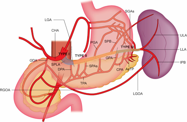
Background: Proximal splenic artery embolisation (PSAE) can be performed in stable patients with Association for the Surgery of Trauma (AAST) grade III-V splenic injury. PSAE reduces splenic perfusion but maintains viability of the spleen and pancreas via the collateral circulation. The hypothesized ideal location is between the dorsal pancreatic artery (DPA) and great pancreatic artery (GPA). This study compares the outcomes resulting from PSAE embolisation in different locations along the splenic artery.

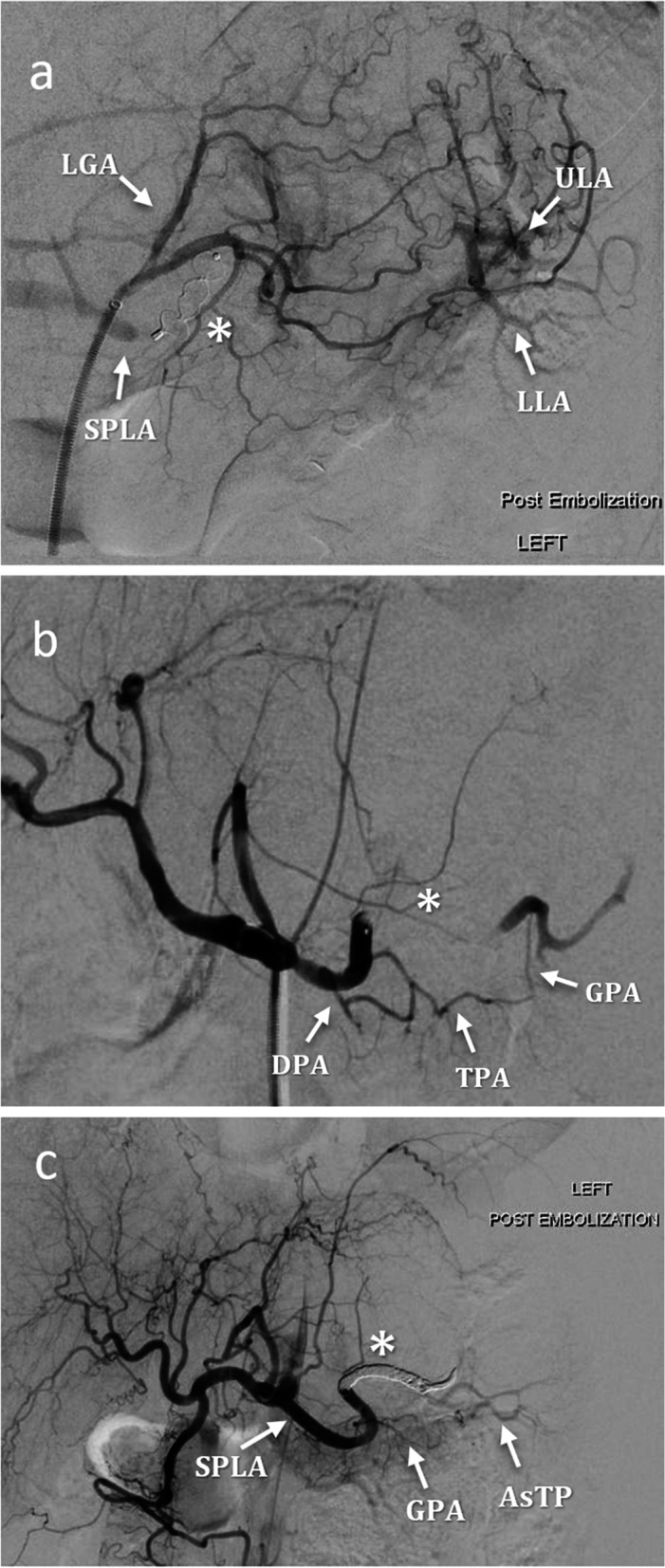
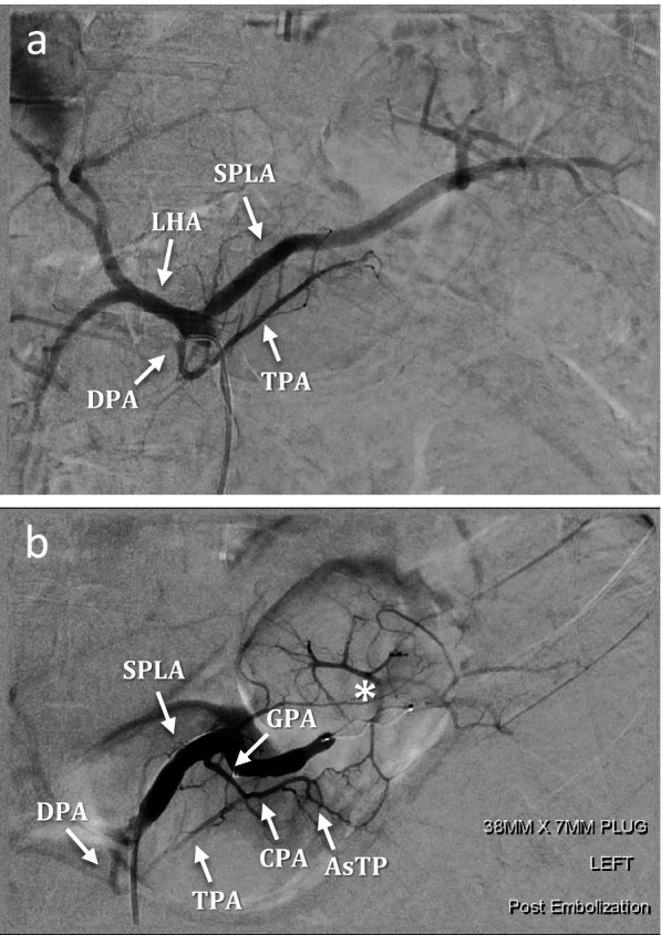
Materials and methods: Retrospective review was performed of PSAE for blunt splenic trauma (2015-2020). Embolisation locations were divided into: Type I, proximal to DPA; Type II, DPA-GPA; Type III, distal to GPA. Fifty-eight patients underwent 59 PSAE: Type I (7); Type II (27); Type III (25). Data was collected on technical and clinical success, post-embolisation pancreatitis and splenic perfusion. Statistical significance was assessed using a chi-squared test.

Results: Technical success was achieved in 100% of cases. Clinical success was 100% for Type I/II embolisation and 88% for Type III: one patient underwent reintervention and two had splenectomies for ongoing instability. Clinical success was significantly higher in Type II embolisation compared to Type III (p = 0.02). No episodes of pancreatitis occurred post-embolisation. Where post-procedural imaging was obtained, splenic perfusion remained 100% in Type I and II embolisation and 94% in Type III. Splenic perfusion was significantly higher in the theorized ideal Type II group compared to Type I and III combined (p = 0.01).

Conclusion: The results support the proposed optimal embolisation location as being between the DPA and GPA.


Conflict of interest statement

The authors declare that they have no competing interests.

Figures

Fig. 1
Fig. 1
Arterial splenic supply. Arteries of the tail of the pancreas (AsTP), common hepatic artery (CHA), caudal pancreatic artery (CPA), great pancreatic artery (GPA), inferior polar branch (IPB), left gastric artery (LGA), left gastro-omental artery (LGOA), lower lobar artery (LLA), posterior gastric artery (PGA), right gastro-omental artery (RGOA), short gastric arteries (SGAs), short pancreatic arteries (SPAs), superior polar Branch (SPB), splenic artery (SPLA), transverse pancreatic artery (TPA), upper lobar artery (ULA)
Fig. 2
Fig. 2
Types of PSAE. a Type I, AVP (white star) was deployed proximally to the DPA, note perfusion of the spleen via prominent LGA collateral circulation. b Type II, AVP (white star) inserted between DPA and GPA origin, spleen perfusion via collateral pancreatic circulation. c Type III, coiling (white star) distal to GPA, minimal splenic perfusion via AsTP
Fig. 3
Fig. 3
PSAE in patient presenting with blunt splenic trauma and variant splenic anatomy. a Pre-embolisation diagnostic angiogram revealed the DPA arising directly from the coeliac trunk along with the left hepatic artery (LHA). CT angiography showed the right hepatic artery origin from the superior mesenteric artery, and the left gastric artery arising from the coeliac trunk, not shown here. b Post-embolisation angiogram shows an AVP plug (white star) inserted distal to the GPA origin and splenic perfusion maintained via the AsTP collateral circulation. This case was thus considered a Type III embolisation

References

    1. Ahuja C, Farsad K, Chadha M. An overview of splenic embolization. AJR Am J Roentgenol. 2015;205(4):720–725. doi: 10.2214/AJR.15.14637. - DOI - PubMed
    1. Anderson JH, VuBan A, Wallace S, Hester JP, Burke JS. Transcatheter splenic arterial occlusion: an experimental study in dogs. Radiology. 1977;125(1):95–102. doi: 10.1148/125.1.95. - DOI - PubMed
    1. Banerjee A, Duane TM, Wilson SP, et al. Trauma center variation in splenic artery embolization and spleen salvage: a multicenter analysis. J Trauma Acute Care Surg. 2013;75(1):69–74. doi: 10.1097/TA.0b013e3182988b3b. - DOI - PubMed
    1. Bessoud B, Denys A. Main splenic artery embolization using coils in blunt splenic injuries: effects on the intrasplenic blood pressure. Eur Radiol. 2004;14(9):1718–1719. doi: 10.1007/s00330-004-2234-3. - DOI - PubMed
    1. Bessoud B, Duchosal MA, Siegrist C-A, et al. Proximal splenic artery embolization for blunt splenic injury: clinical, immunologic, and ultrasound-doppler follow-up. J Trauma Acute Care Surg. 2007;62(6):1481–1486. doi: 10.1097/TA.0b013e318047dfb8. - DOI - PubMed

LinkOut - more resources