Case Reports
. 2022 Aug 20;17(1):192.
doi: 10.1186/s13019-022-01954-z.

Successful removal of the largest reported intrathoracic lipoma with bilateral extension: a case report

Affiliations
Case Reports

Successful removal of the largest reported intrathoracic lipoma with bilateral extension: a case report

S M Tajdit Rahman et al. J Cardiothorac Surg. .

Abstract

Background: Unlike subcutaneous lipomas, thoracic cavity lipomas are extremely rare and can develop to be quite large without causing any symptoms. However, managing massive lipoma that involves both chest cavities is usually challenging, especially when considering the approach for excision.

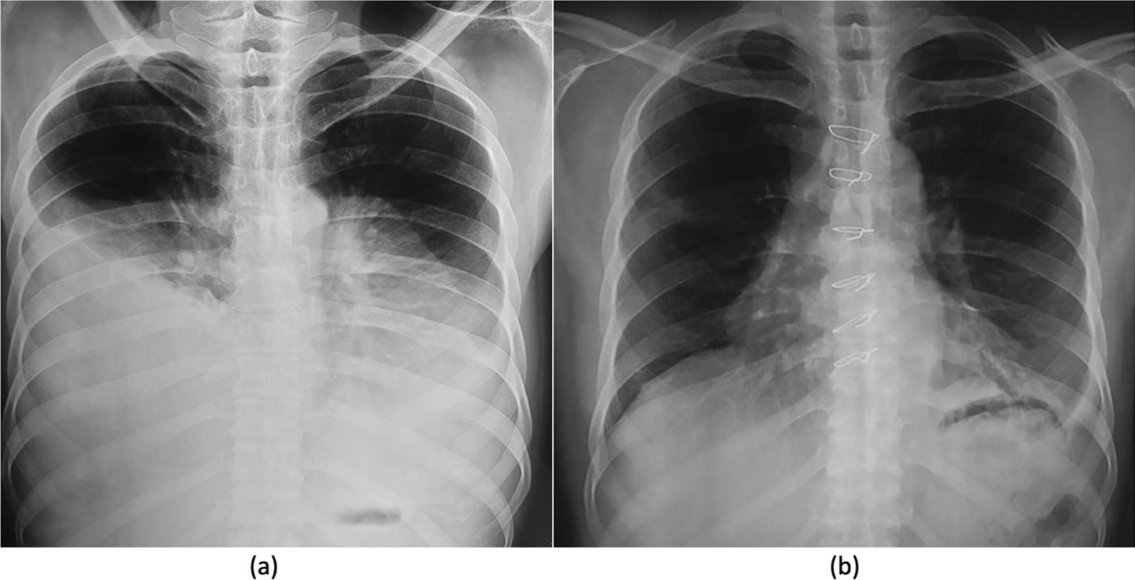
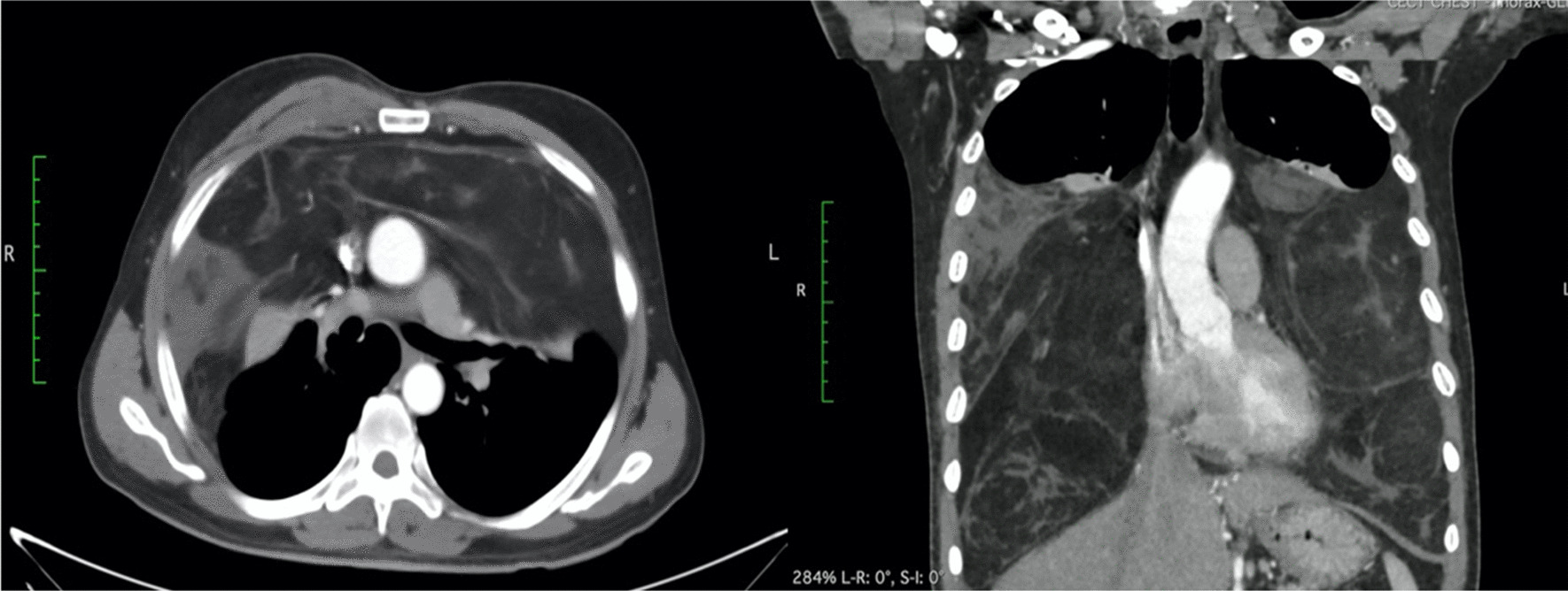
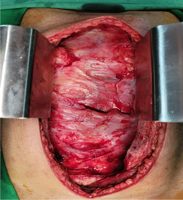
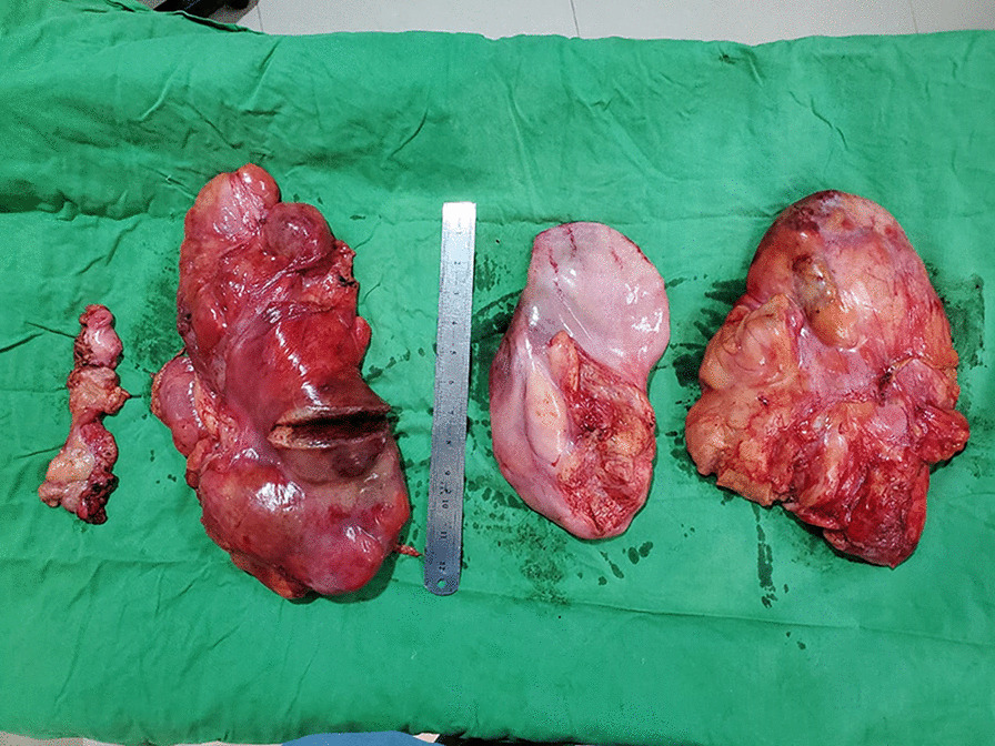
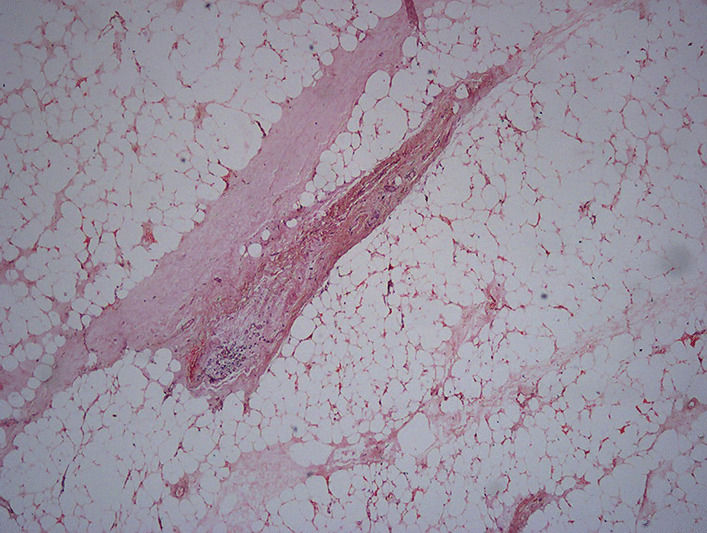
Case: We report our experience of surgical management of a case of a 46-year-old male with huge intrathoracic lipoma that extends bilaterally and is known to be the largest of such kind. The tumor was resected successfully using median sternotomy. Histological analysis confirmed features of lipoma.

Conclusion: To remove a bilateral intrathoracic lipoma, various surgical approaches have been documented. In our experience, a median sternotomy allows better exposure, which aids in complete surgical extirpation resulting in the prevention of recurrence.

Keywords: Bilateral; Case report; Giant; Intrathoracic lipoma; Lipoma; Median sternotomy.


Conflict of interest statement

The authors declare that they have no competing interests.

Figures

Fig. 1
Fig. 1
a Preoperative chest X-ray shows huge mass occupying both hemithorax and displacing both lungs upwards, b postoperative chest X-ray shows no residual mass and well expanded lung after 1 month
Fig. 2
Fig. 2
Contrast CT of the chest shows well-defined fat density mass occupying both hemithorax and displaced both lungs upwards
Fig. 3
Fig. 3
Operative view of the tumor just after median sternotomy
Fig. 4
Fig. 4
The resected specimen shows three large parts of the tumor. Tumors left to the measuring scale were extracted from left side, whereas tumors right to the scale were removed from right chest cavity
Fig. 5
Fig. 5
Microscopic examination reveals abundant mature adipose tissue mixed with fibrous tissue

References

    1. Aldahmashi M, Elmadawy A, Mahdy M, et al. The largest reported intrathoracic lipoma: a case report and current perspectives review. J Cardiothorac Surg. 2019;14:215. doi: 10.1186/s13019-019-1030-8. - DOI - PMC - PubMed
    1. Rahman SMT, Rahim A, Kibria AA. Unusual cause of large intrathoracic mass in a young male of Bangladesh: a case report of giant intrathoracic lipoma and literature review. Int J Surg Case Rep. 2020;76:73–76. doi: 10.1016/j.ijscr.2020.09.164. - DOI - PMC - PubMed
    1. Beshay M, Schmid RA. Intrathoracic giant lipoma. Ann Thorac Surg. 2004;78:1850. doi: 10.1016/S0003-4975(03)01186-X. - DOI - PubMed
    1. Politis J, Funahashi A, Gehlsen JA, DeCock D, Stengel BF, Choi H. Intrathoracic lipomas. Report of three cases and review of the literature with emphasis on endobronchial lipoma. J Thorac Cardiovasc Surg. 1979;77:550–556. doi: 10.1016/S0022-5223(19)38260-1. - DOI - PubMed
    1. Kamata S, Ishida I, Suzuki Y, Yamada T, Yaegashi H, Oura H. Intrathoracic fibrolipoma resected using complete thoracoscopic surgery: a case report. J Cardiothorac Surg. 2018;13(1):112. doi: 10.1186/s13019-018-0808-4. - DOI - PMC - PubMed

Publication types

LinkOut - more resources