

Versatile Nano-PROTAC-Induced Epigenetic Reader Degradation for Efficient Lung Cancer Therapy
- PMID: 35988145
- PMCID: PMC9561860
- DOI: 10.1002/advs.202202039
Versatile Nano-PROTAC-Induced Epigenetic Reader Degradation for Efficient Lung Cancer Therapy
Abstract
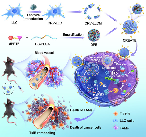
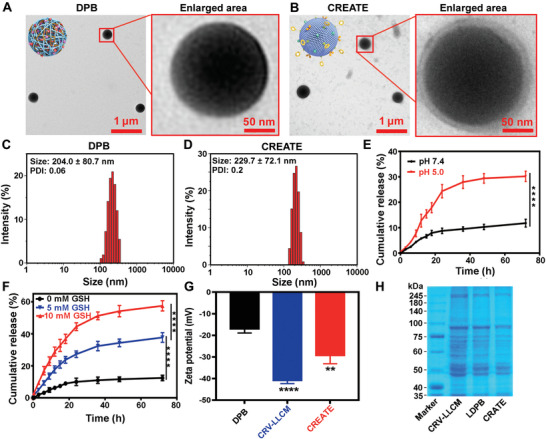
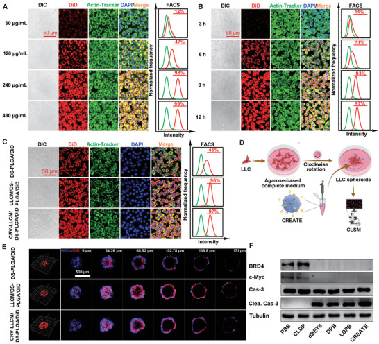
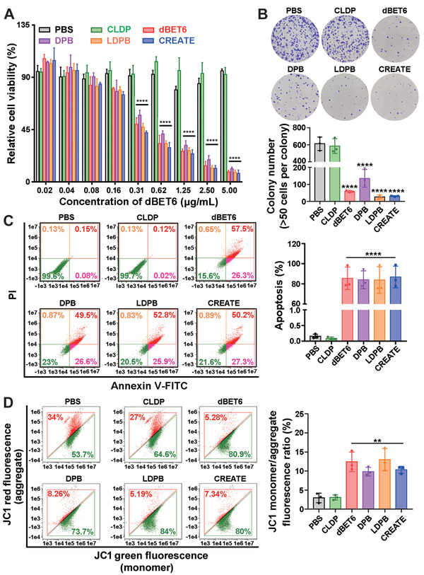
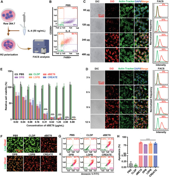
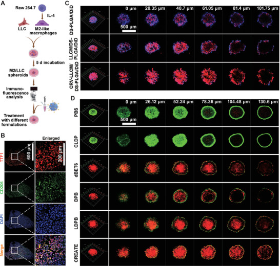
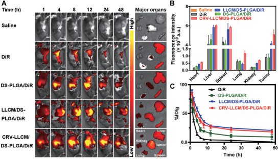
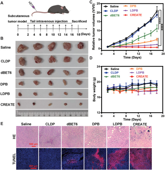
Recent evidence has indicated that overexpression of the epigenetic reader bromodomain-containing protein 4 (BRD4) contributes to a poor prognosis of lung cancers, and the suppression of its expression promotes cell apoptosis and leads to tumor shrinkage. Proteolysis targeting chimera (PROTAC) has recently emerged as a promising therapeutic strategy with the capability to precisely degrade targeted proteins. Herein, a novel style of versatile nano-PROTAC (CREATE (CRV-LLC membrane/DS-PLGA/dBET6)) is developed, which is constructed by using a pH/GSH (glutathione)-responsive polymer (disulfide bond-linked poly(lactic-co-glycolic acid), DS-PLGA) to load BRD4-targeted PROTAC (dBET6), followed by the camouflage with engineered lung cancer cell membranes with dual targeting capability. Notably, CREATE remarkably confers simultaneous targeting ability to lung cancer cells and tumor-associated macrophages (TAMs). The pH/GSH-responsive design improves the release of dBET6 payload from nanoparticles to induce pronounced apoptosis of both cells, which synergistically inhibits tumor growth in both subcutaneous and orthotopic tumor-bearing mouse model. Furthermore, the efficient tumor inhibition is due to the direct elimination of lung cancer cells and TAMs, which remodels the tumor microenvironment. Taken together, the results elucidate the construction of a versatile nano-PROTAC enables to eliminate both lung cancer cells and TAMs, which opens a new avenue for efficient lung cancer therapy via PROTAC.
Keywords: BRD4; PROTAC; epigenetic reader; tumor microenvironment; tumor-associated macrophages.
© 2022 The Authors. Advanced Science published by Wiley-VCH GmbH.
Conflict of interest statement
The authors declare no conflict of interest.
Figures
Similar articles
-
Dual targeting and bioresponsive nano-PROTAC induced precise and effective lung cancer therapy.J Nanobiotechnology. 2024 Nov 10;22(1):692. doi: 10.1186/s12951-024-02967-7. J Nanobiotechnology. 2024. PMID: 39523308 Free PMC article.
-
Tumor microenvironment responsive nano-PROTAC for BRD4 degradation enhanced cancer photo-immunotherapy.Biomaterials. 2025 Nov;322:123387. doi: 10.1016/j.biomaterials.2025.123387. Epub 2025 May 7. Biomaterials. 2025. PMID: 40344878
-
Selective degradation of BRD4 suppresses lung cancer cell proliferation using GSH-responsive PROTAC precursors.Bioorg Chem. 2023 Nov;140:106793. doi: 10.1016/j.bioorg.2023.106793. Epub 2023 Sep 3. Bioorg Chem. 2023. PMID: 37683536
-
Recent progress in degradation of membrane proteins by PROTACs and alternative targeted protein degradation techniques.Eur J Med Chem. 2023 Dec 15;262:115911. doi: 10.1016/j.ejmech.2023.115911. Epub 2023 Oct 30. Eur J Med Chem. 2023. PMID: 37924709 Review.
-
Small-molecule PROTAC degraders of the Bromodomain and Extra Terminal (BET) proteins - A review.Drug Discov Today Technol. 2019 Apr;31:43-51. doi: 10.1016/j.ddtec.2019.04.001. Epub 2019 May 1. Drug Discov Today Technol. 2019. PMID: 31200858 Review.
Cited by
-
The Advances in the Development of Epigenetic Modifications Therapeutic Drugs Delivery Systems.Int J Nanomedicine. 2024 Oct 19;19:10623-10637. doi: 10.2147/IJN.S480095. eCollection 2024. Int J Nanomedicine. 2024. PMID: 39445155 Free PMC article. Review.
-
Cell Death Pathway Regulation by Functional Nanomedicines for Robust Antitumor Immunity.Adv Sci (Weinh). 2024 Jan;11(3):e2306580. doi: 10.1002/advs.202306580. Epub 2023 Nov 20. Adv Sci (Weinh). 2024. PMID: 37984863 Free PMC article. Review.
-
Polymer-Based Drug Delivery Systems for Cancer Therapeutics.Polymers (Basel). 2024 Mar 19;16(6):843. doi: 10.3390/polym16060843. Polymers (Basel). 2024. PMID: 38543448 Free PMC article. Review.
-
PROTAC Delivery Strategies for Overcoming Physicochemical Properties and Physiological Barriers in Targeted Protein Degradation.Pharmaceutics. 2025 Apr 9;17(4):501. doi: 10.3390/pharmaceutics17040501. Pharmaceutics. 2025. PMID: 40284496 Free PMC article. Review.
-
DUSP5 regulated by YTHDF1-mediated m6A modification promotes epithelial-mesenchymal transition and EGFR-TKI resistance via the TGF-β/Smad signaling pathway in lung adenocarcinoma.Cancer Cell Int. 2024 Jun 13;24(1):208. doi: 10.1186/s12935-024-03382-6. Cancer Cell Int. 2024. PMID: 38872157 Free PMC article.
References
-
- Binnewies M., Roberts E. W., Kersten K., Chan V., Fearon D. F., Merad M., Coussens L. M., Gabrilovich D. I., Ostrand‐Rosenberg S., Hedrick C. C., Vonderheide R. H., Pittet M. J., Jain R. K., Zou W., Howcroft T. K., Woodhouse E. C., Weinberg R. A., Krummel M. F., Nat. Med. 2018, 24, 541. - PMC - PubMed
-
- Zheng Y., Shen W., Zhang J., Yang B., Liu Y. N., Qi H., Yu X., Lul S. Y., Chen Y., Xu Y. Z., Lp Y., Gage F. H., Mi S., Yao J., Nat. Neurosci. 2018, 21, 447. - PubMed
Publication types
MeSH terms
Substances
Grants and funding
- 82072470/National Natural Science Foundation of China
- 81871809/National Natural Science Foundation of China
- 81700382/National Natural Science Foundation of China
- 81602360/National Natural Science Foundation of China
- 81672224/National Natural Science Foundation of China
- 2021A1515012154/Natural Science Foundation of Guangdong Province
- 2019A1515011082/Natural Science Foundation of Guangdong Province
- 2019A1515012166/Natural Science Foundation of Guangdong Province
- 2017A030313665/Natural Science Foundation of Guangdong Province
- 202102010069/Guangzhou Science and Technology Project
- 201707010493/Guangzhou Science and Technology Project
- 0029/2019/A/Macau Foundation for Development of Science and Technology
- X20200301018/Youth Talent Support Project of Guangzhou Association for Science & Technology
LinkOut - more resources
Full Text Sources
Medical