. 2022 Sep 14;30(9):1242-1254.e6.
doi: 10.1016/j.chom.2022.07.016. Epub 2022 Aug 4.

Antibody escape and cryptic cross-domain stabilization in the SARS-CoV-2 Omicron spike protein

Affiliations

Antibody escape and cryptic cross-domain stabilization in the SARS-CoV-2 Omicron spike protein

Kamyab Javanmardi et al. Cell Host Microbe. .

Abstract

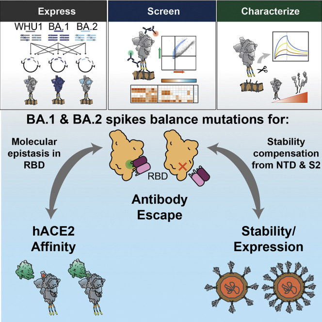
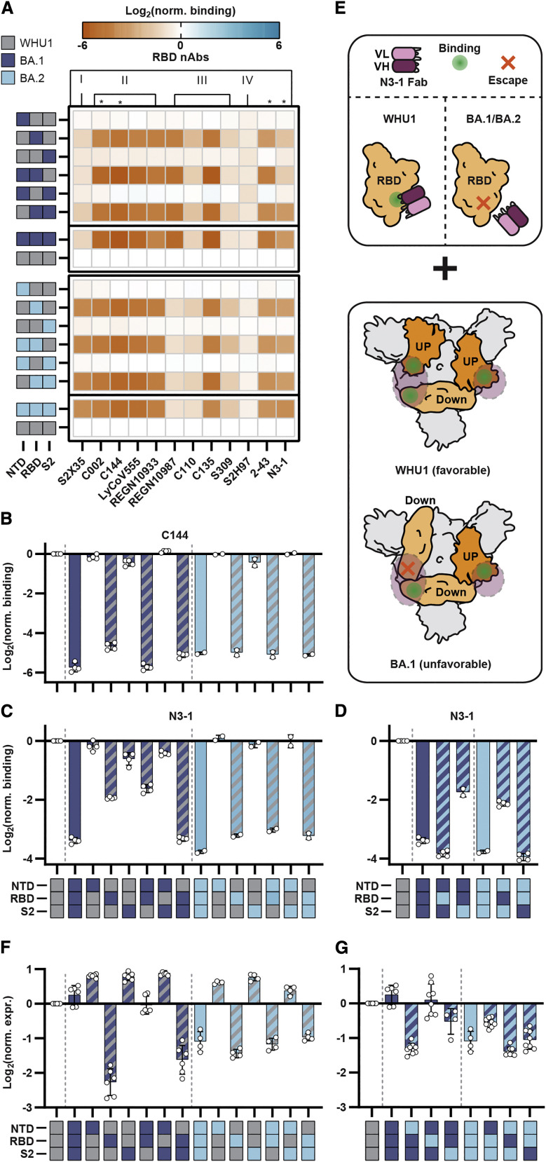
The worldwide spread of severe acute respiratory syndrome coronavirus 2 (SARS-CoV-2) has led to the repeated emergence of variants of concern. For the Omicron variant, sub-lineages BA.1 and BA.2, respectively, contain 33 and 29 nonsynonymous and indel spike protein mutations. These amino acid substitutions and indels are implicated in increased transmissibility and enhanced immune evasion. By reverting individual spike mutations of BA.1 or BA.2, we characterize the molecular effects of the Omicron spike mutations on expression, ACE2 receptor affinity, and neutralizing antibody recognition. We identified key mutations enabling escape from neutralizing antibodies at a variety of epitopes. Stabilizing mutations in the N-terminal and S2 domains of the spike protein can compensate for destabilizing mutations in the receptor binding domain, enabling the record number of mutations in Omicron. Our results provide a comprehensive account of the mutational effects in the Omicron spike protein and illustrate previously uncharacterized mechanisms of host evasion.

Keywords: COVID-19; VOCs; cell display; flow cytometry; high throughput; viral glycoprotein.


Conflict of interest statement

Declaration of interests D.R.B., A.D.E., and J.D.G. have filed patent applications monoclonal antibodies targeting SARS-CoV-2. K.J., C.-W.C., and I.J.F. have filed patent applications on spike 6p (HexaPro).

Figures

None
Graphical abstract
Figure 1
Figure 1
Omicron spike proteins have dozens of mutations contributing antibody escape (A) The number of mutations (aa) for each VOI and VOC. Spike NTD mutations, light blue; spike RBD mutations, yellow; spike S2 mutations, dark blue; other mutations, white. (B) Distribution of all nonsynonymous mutations (substitutions = 42,077,816; insertions = 31,063; deletions = 15,664,146, colored as in A) found in GISAID (accessed on 18/December/2021). The NTD has the most insertions and deletions (81% and 84%, respectively). (C) SARS-CoV-2 spike ectodomain structure (PDB: 7DDN; Zhang et al., 2021) with mutations found in BA.1 and BA.2 colored by domain as in (A). (D) Mutations found in the Omicron spike variants. Shading indicates the percentage of BA.1 or BA.2 strains containing these mutations, as analyzed on outbreak.info (accessed on 12/February/2022). (E) Spike display platform overview. Spike protein ectodomains are constructed using a semi-automated cloning pipeline, displayed on the surface of HEK293T cells, and assayed with flow cytometry. Biophysical characterization is performed with spikes cleaved from cell surfaces. (F) Relative mAb binding to spikes from six VOCs and a VOI. Red, decreased binding; blue, increased binding; normalized to the original WHU1 spike with D614G mutation (top row). Mean ± SD of log-transformed values from at least two biological replicates. Spike domain targets and epitope classifications of antibodies shown on top, , quaternary binding. (G and H) Authentic BA.1 virus neutralization for selected NTD- (G) and RBD-directed mAbs. Mean ± SD of two biological replicates. Curves are a sigmoidal (4PL, X), least squares fit, IC50 values listed in Figure S1E.
Figure 2
Figure 2
NTD indels and substitutions enable mAb binding escape (A) An enlarged NTD structure (PDB: 7DDN; Zhang et al., 2021) with nonsynonymous mutations from BA.1 (brown), BA.2 (gray), or both (black) indicated. (B and C) Relative mAb binding to WHU1 spike proteins containing BA.1-NTD (B) or BA.2-NTD (C) mutations. Red, decreased binding; blue, increased binding; normalized to the WHU1 spike. Mutations color coded as in (A). (D and E) Relative mAb binding to BA.1 (D) or BA.2 (E) spike proteins containing reversions of the respective BA.1- or BA.2-NTD mutations to the WHU1 sequence. Colored as in B, normalized to the BA.1 (D) or BA.2 (E) spike. (F) Comparison of log2(normalized binding) measurements for adding (+1) BA.1-NTD mutations to the WHU1 spike versus reverting (−1) the corresponding mutations from the BA.1 spike protein. Mutations with equal antigenic effects in both spike contexts are expected to fall on the diagonal line y = −x. (G) Comparison of the effect of adding and reverting BA.2-NTD mutations as in (F). For all plots, mean ± SD of log-transformed values from at least two biological replicates.
Figure 3
Figure 3
Omicron-RBD mutations enable antibody evasion and preserve hACE2 affinity (A) An enlarged RBD structure (PDB: 7DDN; Zhang et al., 2021) with mutations in BA.1 (brown), BA.2 (gray), or both (black) indicated. (B) Relative monoclonal antibody binding to WHU1 spike proteins containing BA.1- and BA.2-RBD mutations. Red, decreased binding; blue, increased binding; relative to the WHU1 spike. (C and D) Relative monoclonal antibody binding to BA.1 (C) or BA.2 (D) spike proteins containing reversions of the respective BA.1- or BA.2-RBD mutations to the WHU1 sequence. Colored as in B, normalized to the BA.1 (C) or BA.2 (D) spike. (E) Comparison of the effect of adding (+1) BA.1-RBD mutations to the WHU1 spike protein versus reverting (−1) the corresponding mutations from the BA.1 spike. Mutations with equal antigenic effects in both spike contexts are expected to fall on the diagonal line y = −x. (F) Comparison of the effect of adding and reverting BA.2-RBD mutations as in (E). (G and H) Relative mAb C144 (G) or C135 (H) binding to the WHU1, BA.1, and BA.2 spike proteins and spike proteins containing the indicated substitutions or reversions as appropriate, normalized to the level of binding to the WHU1 spike. (I) Relative monomeric hACE2 binding to WHU1 spikes containing BA.1- and BA.2-RBD mutations (WHU1 base), and BA.1/BA.2 spike proteins containing reversions of the BA.1- or BA.2-RBD mutations (BA.1 base/BA.2 base). Red, decreased binding; blue, increased binding; relative to the WHU1, BA.1, and BA.2 spike proteins as appropriate. (J) Monomeric hACE2 binding to WHU1 and BA.1 spike proteins and variants containing substitutions or reversions as appropriate, measured by BLI. All values are normalized to the binding of the WHU1 spike. For all plots, mean ± SD of log-transformed values from at least two biological replicates.
Figure 4
Figure 4
Cross-domain interactions contribute to mAb escape and stabilize the Omicron spike protein (A) Differences in monoclonal antibody binding of RBD-directed antibodies against spike proteins containing combinations of the NTD, RBD, and S2 mutation sets from the BA.1 and BA.2 variants. Red, decreased binding; blue, increased binding, relative to the WHU1 spike. (B and C) Relative mAb C144 (B) or N3-1 (C) binding to spike proteins containing combinations of the NTD, RBD, and S2 BA.1 and BA.2 mutation sets relative to WHU1. (D) Additional N3-1 binding data as in (C). (E) Proposed escape mechanism for biparatropic antibody (N3-1) by Omicron spike proteins. Antibody binding reduced via mutations at the N3-1 binding epitope on the RBD (top). Top-down view of spike protein trimers with annotated RBD up versus down positions (bottom). Violet circles, N3-1 binding epitope. (F) Differences in expression of spike proteins containing combinations of the NTD, RBD, and S2 BA.1 and BA.2 mutation sets. Data normalized to WHU1 spike expression. (G) Additional expression data as in (F). For all plots, mean ± SD of log-transformed values from at least two biological replicates.

Similar articles

Cited by

References

    1. Barnes C.O., Jette C.A., Abernathy M.E., Dam K.-M.A., Esswein S.R., Gristick H.B., Malyutin A.G., Sharaf N.G., Huey-Tubman K.E., Lee Y.E., et al. SARS-CoV-2 neutralizing antibody structures inform therapeutic strategies. Nature. 2020;588:682–687. doi: 10.1038/s41586-020-2852-1. - DOI - PMC - PubMed
    1. Cameroni E., Bowen J.E., Rosen L.E., Saliba C., Zepeda S.K., Culap K., Pinto D., VanBlargan L.A., De Marco A., di Iulio J., et al. Broadly neutralizing antibodies overcome SARS-CoV-2 Omicron antigenic shift. Nature. 2022;602:664–670. doi: 10.1038/s41586-021-04386-2. - DOI - PMC - PubMed
    1. Cao L., Goreshnik I., Coventry B., Case J.B., Miller L., Kozodoy L., Chen R.E., Carter L., Walls A.C., Park Y.-J., et al. De novo design of picomolar SARS-CoV-2 miniprotein inhibitors. Science. 2020;370:426–431. doi: 10.1126/science.abd9909. - DOI - PMC - PubMed
    1. Cao Y., Wang J., Jian F., Xiao T., Song W., Yisimayi A., Huang W., Li Q., Wang P., An R., et al. Omicron escapes the majority of existing SARS-CoV-2 neutralizing antibodies. Nature. 2022;602:657–663. doi: 10.1038/s41586-021-04385-3. - DOI - PMC - PubMed
    1. Cerutti G., Guo Y., Liu L., Liu L., Zhang Z., Luo Y., Huang Y., Wang H.H., Ho D.D., Sheng Z., Shapiro L. Cryo-EM structure of the SARS-CoV-2 Omicron spike. Cell Rep. 2022;38 doi: 10.1016/j.celrep.2022.110428. - DOI - PMC - PubMed

Supplementary concepts