. 2022 Sep;298(9):102400.
doi: 10.1016/j.jbc.2022.102400. Epub 2022 Aug 18.

Examination of differential glycoprotein preferences of N-acetylglucosaminyltransferase-IV isozymes a and b

Affiliations

Examination of differential glycoprotein preferences of N-acetylglucosaminyltransferase-IV isozymes a and b

Naoko Osada et al. J Biol Chem. 2022 Sep.

Abstract

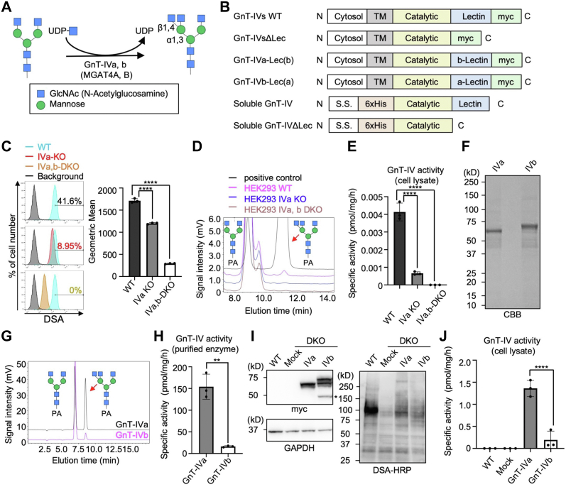
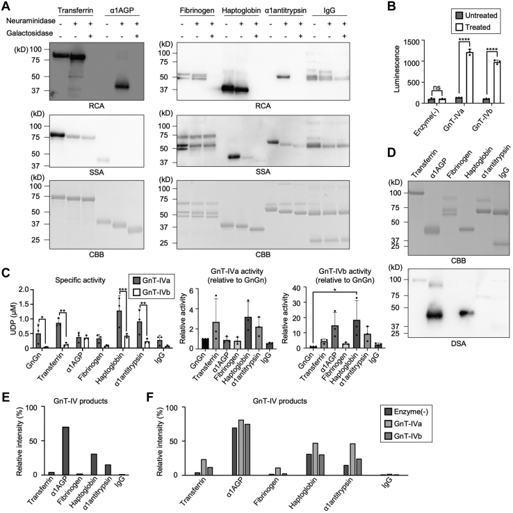
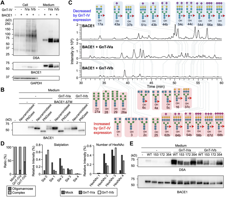
The N-glycans attached to proteins contain various GlcNAc branches, the aberrant formation of which correlates with various diseases. N-Acetylglucosaminyltransferase-IVa (GnT-IVa or MGAT4A) and Gnt-IVb (or MGAT4B) are isoenzymes that catalyze the formation of the β1,4-GlcNAc branch in N-glycans. However, the functional differences between these isozymes remain unresolved. Here, using cellular and UDP-Glo enzyme assays, we discovered that GnT-IVa and GnT-IVb have distinct glycoprotein preferences both in cells and in vitro. Notably, we show that GnT-IVb acted efficiently on glycoproteins bearing an N-glycan premodified by GnT-IV. To further understand the mechanism of this reaction, we focused on the noncatalytic C-terminal lectin domain, which selectively recognizes the product glycans. Replacement of a nonconserved amino acid in the GnT-IVb lectin domain with the corresponding residue in GnT-IVa altered the glycoprotein preference of GnT-IVb to resemble that of GnT-IVa. Our findings demonstrate that the C-terminal lectin domain regulates differential substrate selectivity of GnT-IVa and GnT-IVb, highlighting a new mechanism by which N-glycan branches are formed on glycoproteins.

Keywords: GnT-IV; glycobiology; glycoprotein biosynthesis; glycosylation; glycosyltransferase; substrate specificity.


Conflict of interest statement

Conflict of interest The authors declare that they have no conflicts of interest with the contents of this article.

Figures

Figure 1
Figure 1
In vitro and intracellular activity of GnT-IVa and GnT-IVb.A, schematic model of the GlcNAc transfer reaction catalyzed by GnT-IVa and GnT-IVb (MGAT4A and MGAT4B). B, plasmid constructs used in this study. C, FACS analysis of HEK293 WT (blue), HEK293 GnT-IVa KO (red), and HEK293 DKO cells (orange) with DSA lectin (left). The right graph shows the geometric means (n = 3, means ± SD, ∗∗∗∗p < 0.0001, Tukey's multiple comparisons test). D, the endogenous GnT-IV activity in HEK293 WT, GnT-IVa KO, and DKO cells was measured by incubating the lysates with the PA-labeled acceptor sugar (GnGnbi-PA) and analyzing by HPLC. E, the specific activity of GnT-IV in the HEK293 WT, GnT-IVa KO, and DKO cell lysates calculated by the peak area in (D) (n = 3, means ± SD, ∗∗∗∗p < 0.0001, Tukey's multiple comparisons test). F, soluble GnT-IVa and GnT-IVb were expressed in COS7 cells and purified from the media using a Ni2+ column. Purified GnT-IVa and GnT-IVb were separated by SDS-PAGE and visualized by CBB staining. G, enzyme activity of the purified GnT-IVa and GnT-IVb was measured by incubating the enzymes with GnGnbi-PA and analyzing by HPLC. H, the specific activity of the purified GnT-IVa and GnT-IVb was calculated by the peak area in (G) (n = 3, means ± SD, ∗∗p < 0.01, unpaired t test). I, proteins from mock-treated HEK293 WT cells and DKO cells transfected with an empty vector (mock) or a plasmid for expression of GnT-IVa or GnT-IVb were subjected to SDS-PAGE and blotted with anti-myc antibody (upper left), anti-GAPDH antibody (lower left), or HRP-conjugated DSA (right). J, lysates of mock-treated HEK293 WT cells and DKO cells transfected with an empty vector (mock) or a plasmid for expression of GnT-IVa or GnT-IVb were reacted with GnGnbi-PA and analyzed by HPLC (n = 3, means ± SD, ∗∗∗∗p < 0.0001, Tukey's multiple comparisons test). CBB, Coomassie brilliant blue; DKO, double KO; DSA, Datura stramonium agglutinin; FACS, fluorescence-activated cell sorting; GnT, N-acetylglucosaminyltransferase; HEK293, human embryonic kidney 293 cell line; HRP, horseradish peroxidase.
Figure 2
Figure 2
Intracellular localization of GnT-IVa and GnT-IVb. DKO cells transfected with a plasmid for expression of GnT-IVa or GnT-IVb were stained with anti-myc (green), antibodies for marker proteins (Golgin-97 for the Golgi, calnexin for the ER, or N-cadherin for the plasma membrane) (red), and DAPI (blue). The scale bar represents 10 μm. DAPI, 4′,6-diamidino-2-phenylindole; DKO, double KO; ER, endoplasmic reticulum; GnT, N-acetylglucosaminyltransferase.
Figure 3
Figure 3
The in vitro activity of soluble GnT-IVa and GnT-IVb toward an oligosaccharide and various glycoproteins.A, glycoprotein substrates for the UDP-Glo assay were pretreated with neuraminidase and β-galactosidase. The treated proteins were stained with RCA, SSA, or CBB. B, the UDP-Glo assay was performed using purified soluble GnT-IVa or GnT-IVb and glycosidase-treated or untreated α1AGP (n = 3, means ± SD, ∗∗∗∗p < 0.0001; ns, not significant, Holm–Sidak's multiple comparisons test). C, comparison of the in vitro activity of GnT-IVa and GnT-IVb toward an oligosaccharide and various glycoproteins using the UDP-Glo assay (n = 3, means ± SD, ∗p < 0.05, ∗∗p < 0.01, ∗∗∗p < 0.001, Holm–Sidak's multiple comparisons test [left]; n = 3, means ± SD, ∗p < 0.05, Tukey's multiple comparisons test [middle and light]). D, the glycosidase-treated proteins were stained with CBB or DSA. E, sum of the signal intensities of GnT-IV product N-glycans (#2c, 3, 5c, 6, and 11d in Table S2) in the sialidase- and galactosidase-treated six glycoprotein substrates without incubating with GnT-IV from LC–MS analysis. F, the sialidase- and galactosidase-treated six glycoprotein substrates were incubated with GnT-IVa or GnT-IVb in vitro. Sum of the signal intensities of GnT-IV product N-glycans from LC–MS analysis is shown. α1AGP, alpha-1-acid glycoprotein; CBB, Coomassie brilliant blue; DSA, Datura stramonium agglutinin; GnT, N-acetylglucosaminyltransferase; RCA, Ricinus communis agglutinin; SSA, Sambucus sieboldiana agglutinin.
Figure 4
Figure 4
Differential actions of GnT-IVa and GnT-IVb toward BACE1 in cells.A, cell lysates (cell) and secreted BACE1 purified with Ni2+ beads (medium) were analyzed by Western and lectin blotting. Proteins from HEK293 DKO cells transfected with the plasmids for expression of GnT-IVa, GnT-IVb, and BACE1, or empty vector, were subjected to SDS-PAGE and blotted with HRP-conjugated DSA (upper), anti-BACE1 antibody (middle), and anti-GAPDH antibody (lower). B, secreted BACE1 purified with Ni2+ beads was treated with neuraminidase or PNGaseF, subjected to SDS-PAGE, and blotted with the anti-BACE1 antibody. C, LC–MS total ion chromatograms (TICs) of N-glycans derived from secreted BACE1 with or without coexpression of GnT-IVa or GnT-IVb in HEK293 DKO cells. Pink, glycans increased by coexpression with GnT-IVa or GnT-IVb; blue, glycans decreased by coexpression with GnT-IVa or GnT-IVb. D, left, sum of the signal intensities of oligomannose glycans and hybrid or complex glycans from LC–MS analysis is shown. Middle, sum of the signal intensities of non-, mono-, di-, tri-, and tetrasialylated N-glycans from LC–MS analysis is shown. Right, sum of the signal intensities of N-glycans with 1, 2, 3, and 4 HexNAc residues (HexNAc residues in chitobiose were excluded) from LC–MS analysis is shown. E, secreted BACE1 WT and its N-glycosylation site mutants (N153S, N172S, and N354S) purified with Ni2+ beads were subjected to SDS-PAGE and blotted with HRP-conjugated DSA (upper) and the anti-BACE1 antibody (lower). BACE1, β-site amyloid precursor protein cleaving enzyme-1; DKO, double KO; DSA, Datura stramonium agglutinin; GnT, N-acetylglucosaminyltransferase; HEK293, human embryonic kidney 293 cell line; HRP, horseradish peroxidase; PNGaseF, peptide N-glycanase F.
Figure 5
Figure 5
Lectin domain regulates protein selectivity of GnT-IV.A, proteins from mock-treated HEK293 WT and DKO cells transfected with an empty vector (mock) or a plasmid to express myc-tagged GnT-IVa, GnT-IVb, GnT-IVaΔLec, or GnT-IVbΔLec were subjected to SDS-PAGE and blotted with the anti-myc antibody or anti-GAPDH antibody. B, cell lysates of mock-treated HEK293 WT and DKO cells transfected with an empty vector (mock) or a plasmid for expression of GnT-IVa, GnT-IVb, GnT-IVaΔLec, or GnT-IVbΔLec were reacted with GnGnbi-PA and analyzed by HPLC (n = 3, means ± SD, ∗∗p < 0.01, Tukey's multiple comparisons test). C, proteins from mock-treated HEK293 WT and DKO cells transfected with an empty vector (mock) or a plasmid for expression of GnT-IVa, GnT-IVb, GnT-IVa-Lec(b), or GnT-IVb-Lec(a) were subjected to SDS-PAGE and blotted with the anti-myc antibody or anti-GAPDH antibody. D, lysates of mock-treated HEK293 WT and DKO cells transfected with an empty vector (mock) or a plasmid for expression of GnT-IVa, GnT-IVb, GnT-IVa-Lec(b), or GnT-IVb-Lec(a) were reacted with GnGnbi-PA and analyzed by HPLC (n = 3, means ± SD, ∗∗∗p < 0.001, ∗∗∗∗p < 0.0001, Tukey's multiple comparisons test). E, the structure of the lectin domain of GnT-IVa modeled with GlcNAc. The residues conserved between GnT-IVa and GnT-IVb are shown in red. F, soluble GnT-IVb, GnT-IVb F413H, or GnT-IVb I456Q was expressed in COS7 cells and purified from the media using a Ni2+ column. Purified soluble GnT-IVb, GnT-IVb F413H, or GnT-IVb I456Q was separated by SDS-PAGE and visualized by CBB staining. G, the specific activity of the purified GnT-IVb, GnT-IVb F413H, and GnT-IVb I456Q toward GnGnbi-PA was analyzed by HPLC (n = 3, means ± SD, a significant p value was not observed, Tukey's multiple comparisons test). H, comparison of the in vitro activity of GnT-IVb, GnT-IVb F413H, and GnT-IVb I456Q toward an oligosaccharide and various glycoproteins (same as Fig. 3) using the UDP-Glo assay (n = 3, means ± SD, ∗∗p < 0.01, ∗∗∗∗p < 0.0001, Tukey's multiple comparisons test). CBB, Coomassie brilliant blue; DKO, double KO; GnT, N-acetylglucosaminyltransferase; HEK293, human embryonic kidney 293 cell line.

Similar articles

Cited by

References

    1. Varki A. Biological roles of glycans. Glycobiology. 2017;27:3. - PMC - PubMed
    1. Moremen K.W., Tiemeyer M., Nairn A.V. Vertebrate protein glycosylation: diversity, synthesis and function. Nat. Rev. Mol. Cell Biol. 2012;13:448–462. - PMC - PubMed
    1. Zhao Y.-Y., Takahashi M., Gu J.-G., Miyoshi E., Matsumoto A., Kitazume S., et al. Functional roles of N-glycans in cell signaling and cell adhesion in cancer. Cancer Sci. 2008;99:1304–1310. - PMC - PubMed
    1. Mereiter S., Balmaña M., Campos D., Gomes J., Reis C.A. Glycosylation in the era of cancer-targeted therapy: where are we heading? Cancer Cell. 2019;36:6–16. - PubMed
    1. Wang X., Inoue S., Gu J., Miyoshi E., Noda K., Li W., et al. Dysregulation of TGF-beta1 receptor activation leads to abnormal lung development and emphysema-like phenotype in core fucose-deficient mice. Proc. Natl. Acad. Sci. U. S. A. 2005;102:15791–15796. - PMC - PubMed

Publication types

LinkOut - more resources