Review
. 2022 Aug 22;6(1):42.
doi: 10.1186/s41747-022-00291-z.

The potential of predictive and prognostic breast MRI (P2-bMRI)

Affiliations
Review

The potential of predictive and prognostic breast MRI (P2-bMRI)

Matthias Dietzel et al. Eur Radiol Exp. .

Abstract

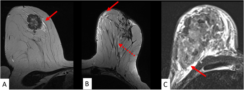
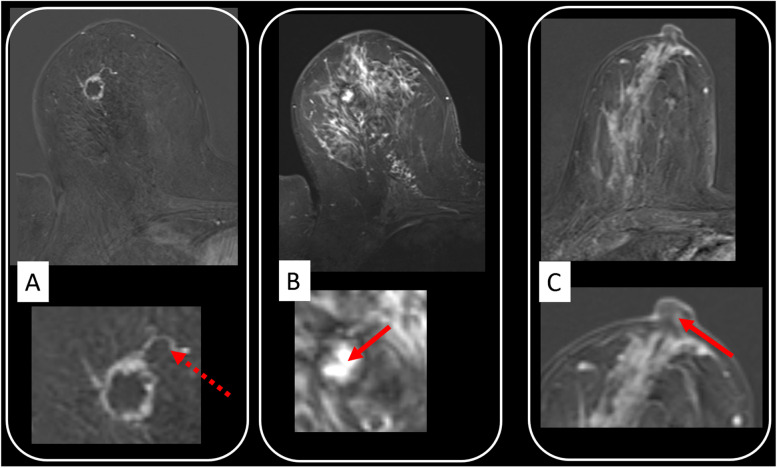
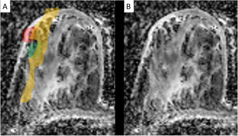
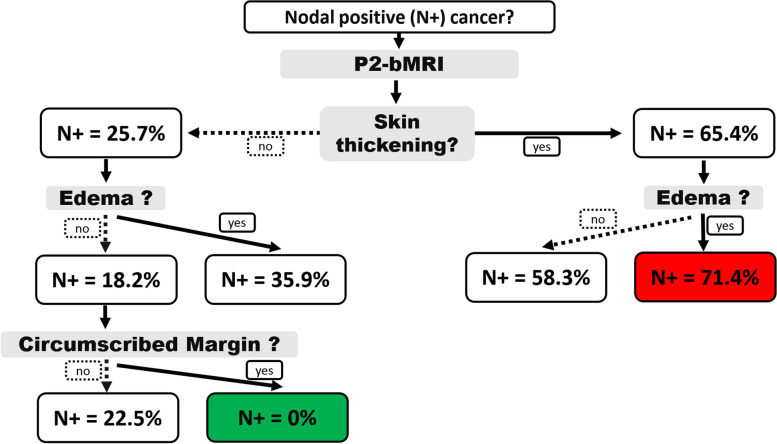
Magnetic resonance imaging (MRI) is an important part of breast cancer diagnosis and multimodal workup. It provides unsurpassed soft tissue contrast to analyse the underlying pathophysiology, and it is adopted for a variety of clinical indications. Predictive and prognostic breast MRI (P2-bMRI) is an emerging application next to these indications. The general objective of P2-bMRI is to provide predictive and/or prognostic biomarkers in order to support personalisation of breast cancer treatment. We believe P2-bMRI has a great clinical potential, thanks to the in vivo examination of the whole tumour and of the surrounding tissue, establishing a link between pathophysiology and response to therapy (prediction) as well as patient outcome (prognostication). The tools used for P2-bMRI cover a wide spectrum: standard and advanced multiparametric pulse sequences; structured reporting criteria (for instance BI-RADS descriptors); artificial intelligence methods, including machine learning (with emphasis on radiomics data analysis); and deep learning that have shown compelling potential for this purpose. P2-bMRI reuses the imaging data of examinations performed in the current practice. Accordingly, P2-bMRI could optimise clinical workflow, enabling cost savings and ultimately improving personalisation of treatment. This review introduces the concept of P2-bMRI, focusing on the clinical application of P2-bMRI by using semantic criteria.

Keywords: Biomarkers; Breast neoplasms; Magnetic resonance imaging; Precision medicine; Prognosis.


Conflict of interest statement

Matthias Dietzel and Pascal Baltzer are members of the European Radiology Experimental Editorial Board. They have not taken part in the review or selection process of this article. Francesco Sardanelli is the Editor in Chief of European Radiology Experimental. He has not taken part in the review or selection process of this article which was fully managed by the Deputy Editor Akos Varga-Szemes (Medical University of South Carolina, Charleston, SC, USA). All remaining authors declare that they have no competing interests.

Figures

Fig. 1
Fig. 1
A 15-min clinical protocol for breast magnetic resonance imaging (MRI). All predictive/prognostic breast MRI information demonstrated in the next figures can be derived from a one-stop shop clinical protocol as shown in this figure. The protocol starts with an unenhanced T2-weighted turbo spin-echo sequence (T2w TSE). Diffusion-weighted imaging (DWI) and short-tau inversion recovery (STIR) are optional but highly recommend. On T2-weighted images, a mass lesion is diagnosed, with perifocal oedema. Next, contrast-enhanced dynamic scanning is performed using a T1-weighed gradient-echo (GRE) sequence before/after the intravenous administration of 0.1 mmol/kg of a Gd-based contrast agent. There is evidence of washout, perifocal oedema, and central necrosis (rim sign). The last two descriptors are imaging biomarkers associated with increased probability of high-grade and nodal-positive invasive cancers. Washout is a strong predictor of poor outcome and is associated with a higher likelihood of metachronous metastasis (see also Figs. 5 and 6). Example taken from ref [2], with permission (Dietzel et al. Insights Imaging 2018)
Fig. 2
Fig. 2
Standardised reading setup for comprehensive diagnostic and predictive/prognostic breast magnetic resonance imaging (MRI) at one-stop shop. A female patient with suspicious amorphous segmental calcifications on the right breast at mammography, breast imaging reporting and data system (BI-RADS) 4 diagnostic category (not shown). MRI was performed also for preoperative staging due to suspicion of extended ductal carcinoma in situ. Diagnostic MRI shows an extensive heterogeneous segmental non-mass enhancement predominantly with plateau dynamic pattern. A noncircumscribed mass with washout and heterogeneous internal enhancement (BI-RADS 5 diagnostic category) is located centrally within the non-mass lesions. Relevant prognostic findings are here washout, skin thickening, invasion of the nipple, and diffuse ipsilateral oedema (see also Figs. 5 and 6). Semantic criteria correspond to the MRI phenotype of an aggressive invasive breast cancer. P2-MRI results were confirmed by postoperative pathological examination (invasive cancer NOS, G3, Ki-67+++, triple negative, node positive). Apparent diffusion coefficient map (A); unenhanced T1-weighted gradient echo with colour overlap of the dynamic curve on a pixel-by-pixel basis (green/yellow/red = persistent/plateau/washout) (B); first (C) and last (D) contrast-enhanced T1 GRE; diffusion-weighted imaging obtained with b = 800 s/mm2 (E); T2-weighted turbo spin-echo (F); first (G) and last (H) contrast-enhanced subtractions. Diffusion-weighted imaging findings are highlighted in Fig. 7
Fig. 3
Fig. 3
Benefit of vascular analysis for predictive/prognostic breast magnetic resonance imaging. In A, B, and C, the enhancement patterns of three different breast cancers are colour coded on a pixel-by-pixel basis (green/yellow/red = persistent/plateau/washout). The maps reveal tumour heterogeneity, progressively increasing from A to C (no washout pixels in A, few of them in B, and many in C). Findings correspond to an increasing risk profile which was verified upon pathological and molecular analysis. Here, increasing vascularisation (CD 31 staining top row), cellular proliferation (Ki-67), and aggressiveness (grading) was demonstrated, and a less favourable receptor profile was evident from A to C. ER Oestrogen, her2neu Human epidermal growth factor receptor 2, PR Progesterone
Fig. 4
Fig. 4
Potential role of T2-weighed images: the lesion signal as a possible surrogate of water content. Signal intensity on T2-weighted images may reflect water content and warrants further scientific research. Compared to the surrounding parenchyma, a lesion is classified as hyperintense (A), isointense (B), or hypointense (C). In D, a mass lesion is identified on T2-weighted images (red arrow). T2-weighted signal intensity is assessed in comparison with the surrounding parenchyma. In D, just like the adjacent Cooper ligaments, the parenchyma displays less signal than the tumour. Correspondingly, the tumour is classified as “hyperintense”. Findings suggest the presence of an aggressive breast carcinoma phenotype. Predictive/prognostic findings were verified by immune histology revealing high-grade cancer with negative steroid receptors and elevated Ki-67 suggesting high cellular proliferation. Note perifocal and subcutaneous oedema (dotted arrows). In contrast, E displays a hypointense less aggressive carcinoma (red arrow, G2, positive steroid receptors, only Ki-67+)
Fig. 5
Fig. 5
The pivotal role of T2-weighed images: oedema. Any asymmetric ipsilateral T2-weighted signal increase not due to the tumour itself, cysts or artefacts is referred to as “oedema” in A, B, and C. Different oedema patterns are distinguished such as perifocal (A), full red arrow), subcutaneous (B, full red arrow), prepectoral (C, full red arrow), and diffuse (B, dotted red arrow). Oedema is considered among the best evaluated predictive/prognostic criteria and was associated with high grade and nodal-positive cancers as well as disease recurrence (see also Table 1)
Fig. 6
Fig. 6
Selected semantic parameters with known biological correlates. They can be readily implemented into clinical practice. Rim enhancement (A) was among the first morphologic parameters reported in the literature and reflects an aggressive cancers phenotype. This is also suggested by the adjacent vessel sign (A, magnification: dotted red arrow) [50]. Rim enhancement is thought to reflect central hypovascularity due to connective tissue, fibrosis, and/or necrosis. Necrosis sign (B) specifically depicts central colliquative (liquid) necrosis (B, magnification: full red arrow), characterised by a high signal intensity on T2-weighted images within the centre of the tumour. Invasion of the cancer into the nipple areolar complex is related to poor outcome. The semantic criterion described as “destruction of nipple line” (C) is best depicted on DCE images (C, magnification: red arrow). Further details including diagnostic performance of semantic parameters are provided in Table 1
Fig. 7
Fig. 7
Apparent diffusion coefficient (ADC) mapping (detailed analysis of the DWI already shown in Fig. 1). ADC values are extracted using quantitative region-of-interest-based measurements by standardised methods. ADC is given with (A) and without (B) colour overlap. ADC was applied to distinguish ductal carcinoma in situ (DCIS, orange) from invasive carcinoma (red). Note the presence of a benign lesion (a fibroadenoma), also correctly characterised by the ADC map (green), adjacent to the DCIS
Fig. 8
Fig. 8
P2-bMRI phenotypes are imaging patterns highly specific of a distinct tumour biology. They may be used as rule-in or rule-out criteria for clinical decision-making. Typically, P2-bMRI phenotypes are based on the assessment of multiple criteria in concert as in this example: here, a machine learning algorithm was used to identify phenotypes predictive of nodal-positive or nodal-negative stage (N+, N-). Semantic imaging criteria of the index lesion were used to predict nodal stage (for details, please see reference [42]). Classification results are presented as an intuitive and easy to follow decision tree. Accordingly, the “nodal-negative P2-bMRI phenotype” is characterised by a smooth lesion without oedema and without skin thickening. The positive likelihood of N+ is 0% for this P2-bMRI phenotype. Similar results can be achieved with other predictive/prognostic MRI methods, including artificial intelligence, each of them providing intrinsic advantages and disadvantages

References

    1. Dietzel M, Baltzer PAT. How to use the Kaiser score as a clinical decision rule for diagnosis in multiparametric breast MRI: a pictorial essay. Insights Imaging. 2018;9:325. doi: 10.1007/s13244-018-0611-8. - DOI - PMC - PubMed
    1. Baltzer PAT, Krug KB, Dietzel M (2022) Evidence-Based and Structured Diagnosis in Breast MRI using the Kaiser Score. Rofo. 10.1055/a-1829-5985. - PubMed
    1. Sardanelli F, Boetes C, Borisch B, et al. Magnetic resonance imaging of the breast: recommendations from the EUSOMA working group. Eur J Cancer. 2010;46:1296–1316. doi: 10.1016/j.ejca.2010.02.015. - DOI - PubMed
    1. Mann RM, Kuhl CK, Kinkel K, Boetes C. Breast MRI: guidelines from the European Society of Breast Imaging. Eur Radiol. 2008;18:1307–1318. doi: 10.1007/s00330-008-0863-7. - DOI - PMC - PubMed
    1. Baltzer P, Mann RM, Iima M, et al. Diffusion-weighted imaging of the breast—a consensus and mission statement from the EUSOBI International Breast Diffusion-Weighted Imaging working group. Eur Radiol. 2019;30:1436–1450. doi: 10.1007/s00330-019-06510-3. - DOI - PMC - PubMed