. 2022 Oct;45(10):1194-1206.
doi: 10.1111/pace.14582. Epub 2022 Sep 1.

Impact of high frequency stimulation to confirm a complete box isolation in catheter ablation of non-paroxysmal atrial fibrillation

Affiliations

Impact of high frequency stimulation to confirm a complete box isolation in catheter ablation of non-paroxysmal atrial fibrillation

Kazuo Kato et al. Pacing Clin Electrophysiol. 2022 Oct.

Abstract

Introduction: Pulmonary vein (PV) isolation (PVI) including the left atrial posterior wall (LAPW) (Box-PVI) is proposed as an additional strategy for non-paroxysmal atrial fibrillation (NPAF), however, the efficacy remains controversial. The more reliable and durable the Box-PVI we can create, the better the rhythm outcomes might be than with a conventional PVI alone. This study focused on the potential exit conduction of the box lesion and investigated whether the conventional Box-PVI would be sufficient.

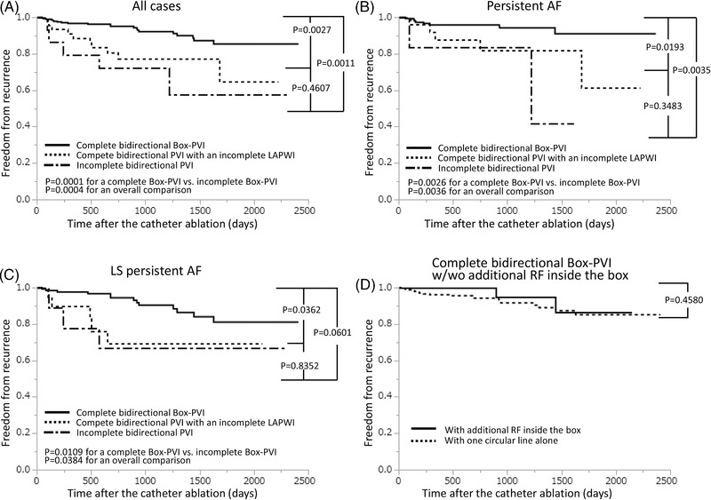
Methods and results: We enrolled 350 consecutive patients with NPAF that underwent a conventional encircling Box-PVI and examined whether latent exit conduction and dormant "exit" conduction independently remained on the LAPW and in the PVs using high frequency stimulation (HFS) and an adenosine triphosphate (ATP) injection. All electrograms inside the box lesion were eliminated in all cases, however, HFS inside the box propagated outward in 23 cases (6.6%) without any exit conduction by conventional burst stimulation, and 24 cases (6.9%) exhibited only dormant "exit" conduction of the LAPW. Additional ablation where positive HFSs were observed created a complete bidirectional Box-PVI in 43 (41.3%) of the cases without a first pass Box-PVI. The recurrence rates depended on the groups classified according to the HFS response.

Conclusion: HFS delivered with an ATP injection on the LAPW and in the PVs following a Box-PVI could not only elucidate true exit block but also identified possible incomplete lesions or connections outside the ablation line, whose elimination could achieve a complete Box-PVI leading to a better rhythm outcome.

Keywords: adenosine triphosphate; box isolation; dormant conduction; exit block; pulmonary vein isolation.


Conflict of interest statement

This research did not receive any specific grant from funding agencies in the public, commercial, or not‐for‐profit sectors. All the authors have no conflicts to disclose.

Figures

FIGURE 1
FIGURE 1
Intracardiac tracings recorded during high frequency stimulation (HFS) on the left atrial posterior wall (LAPW) during sinus rhythm (SR) in the cases before the ablation with a contact force of 1–2 g (A) and 9 g (B), respectively. The atrial rhythm became irregular during the HFS (A), and the HFS disrupted the atrial rhythm provoking AF after cessation of the HFS (B). We defined those as an HFS (+) response. The tracings are surface ECG leads I, aVF, V1, V5, and the intracardiac electrograms recorded by the distal to proximal electrodes of the high right atrial (HRAd‐p) catheter, distal to proximal electrograms recorded by a circular catheter within the pulmonary vein (PVd‐p), distal, proximal, and unipolar electrograms recorded by an ablation catheter (ABLd, ABLp, and ABLu) on the LAPW, distal to proximal coronary sinus catheter (CSd‐p) electrograms, and the blood pressure.
FIGURE 2
FIGURE 2
Flowchart of the procedure and outcomes. Conventional entrance block of the box lesion was obtained in all study cases, however, the response of the HFS and conventional burst stimulation (CBS) differed among the individuals. Moreover, a concomitant adenosine triphosphate (ATP) injection could elicit dormant “exit” conduction in addition to the dormant “entrance” conduction suggesting incomplete lesions. The data are presented as the n (%). See the text and Table 2 for the details. AF indicates atrial fibrillation; Box‐PVI, box pulmonary vein (PV) isolation, LAPWI, LAPW isolation; PVI, PV isolation; RF, radiofrequency energy application. The other abbreviations are the same as in Figure 1.
FIGURE 3
FIGURE 3
Intracardiac tracings recorded during the HFS on the LAPW during SR in the cases after a complete Box‐PVI (A) and incomplete Box‐PVI (B). A: Bump artifact (asterisks) of the catheter on the LAPW that preceded the PV electrograms did not conduct to the atrium, which certified that the catheter was in contact with the LAPW, and the atrial rhythm was not affected during or after the HFS. We defined this tracing response as an HFS (‐) response. B: The atrial rhythm was affected during the HFS, which was an HFS (+) response. The order of the tracings and abbreviations are the same as in Figures 1 and 2.
FIGURE 4
FIGURE 4
Intracardiac tracings recorded in the cases with an incomplete Box‐PVI. (A) The CBS on the LAPW did not propagate outward resulting in maintaining SR, that is, a CBS (‐) response, however, the following HFS disrupted the SR, that is, an HFS (+) response. (B) The atrial rhythm was affected during the HFS, which was an HFS (+) response, however, automatic firing was frequently observed. The order of the tracings and abbreviations are the same as in Figures 1 and 2.
FIGURE 5
FIGURE 5
Intracardiac tracings recorded during the HFS on the LAPW during SR while provoking atrio‐ventricular block by an ATP administration after a Box‐PVI in the cases that underwent a complete Box‐PVI (A) and incomplete Box‐PVI (B). A: The atrial rhythm was not affected by the HFS, which represented an HFS (‐) response. Note that the bump artifact (asterisks) of the catheter on the LAPW was similar to that in Figure 3A, which certified that the catheter was in contact with the LAPW. B: The atrial rhythm was affected by the HFS, which was an HFS (+) response, that is, dormant “exit” conduction, however, non‐conducted automatic firing before and after the HFS was observed. The order of the tracings and abbreviations are the same as in Figures 1 and 2.
FIGURE 6
FIGURE 6
Kaplan‐Meier curves of the comparison of the freedom from atrial tachyarrhythmias after a Box‐PVI among the different isolated levels classified by the HFS response in all patients (A), that in patients with persistent AF (B), that in patients with long‐standing (LS) persistent AF (C), and that in all patients in which a complete bidirectional Box‐PVI was performed between that with or without additional RF inside the box lesion (D). See the text for the details. The abbreviations are the same as in Figures 1 through 2.

References

    1. Kumar P, Bamimore AM, Schwartz JD, et al. Challenges and outcomes of posterior wall isolation for ablation of atrial fibrillation. J Am Heart Assoc. 2016;5:e003885. doi: 10.1161/JAHA.116.003885 - DOI - PMC - PubMed
    1. Thiyagarajah A, Kadhim K, Lau DH, et al. Feasibility, safety, and efficacy of posterior wall isolation during atrial fibrillation ablation. Circ Arrhythm Electrophysiol. 2019;12:e007005. doi: 10.1161/CIRCEP.118.007005 - DOI - PubMed
    1. Markman TM, Hyman MC, Kumareswaran R, et al. Durability of posterior wall isolation after catheter ablation among patients with recurrent atrial fibrillation. Hear Rhythm. 2021;17:1740‐1744. doi: 10.1016/j.hrthm.2020.05.005 - DOI - PubMed
    1. Takigawa M, Derval N, Martin CA, et al. Mechanism of recurrence of atrial tachycardia: comparison between first versus redo procedures in a high‐resolution mapping system. Circ Arrhythm Electrophysiol. 2020;13:e007273. doi: 10.1161/CIRCEP.119.007273 - DOI - PubMed
    1. Haïssaguerre M, Jaïs P, Shah DC, et al. Spontaneous initiation of atrial fibrillation by ectopic beats originating in the pulmonary veins. N Engl J Med. 1998;339:659‐666 - PubMed