Case Reports
. 2022 Jul 19;14(7):e27043.
doi: 10.7759/cureus.27043. eCollection 2022 Jul.

A Rare Case of COVID-19 Infection Leading to Colonic Stricture: Case Report and Review of Literature

Affiliations
Case Reports

A Rare Case of COVID-19 Infection Leading to Colonic Stricture: Case Report and Review of Literature

Komal Yousaf et al. Cureus. .

Abstract

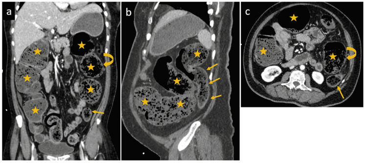
Coronavirus disease 2019 (COVID-19) predominantly targets the respiratory tract; despite gastrointestinal (GI) symptoms that may present in many patients, colonic strictures in coronavirus disease (COVID-19) patients are extremely rare and, to our knowledge, have never been reported. We, herein, present a case of a 59-year-old lady who developed intestinal obstruction due to colonic strictures shortly after recovering from complicated COVID-19 pneumonia. Ultimately, she underwent laparoscopic subtotal colectomy with ileorectal anastomosis. After a long recovery period, she was discharged in good status. It has been more than two years since COVID-19 was declared as a pandemic by the World Health Organization. Infected individuals have highly variable clinical manifestations, yet the pathogenesis, diagnosis and ideal management of each of these complications is not well described in literature.

Keywords: colon; constipation; covid-19; intestinal obstruction; sars-cov-2; strictures.


Conflict of interest statement

The authors have declared that no competing interests exist.

Figures

Figure 1
Figure 1. Frontal chest X-ray shows bilateral multiple confluent alveolar peripheral and medial peribronchovascular infiltrations with air bronchogram.
Figure 2
Figure 2. Initial contrast-enhanced abdominal CT scan.
Coronal (A & B) and axial (C) images show diffuse dilation of the cecum, ascending and transverse colon (stars) up to the splenic flexure, followed by smooth tapering into the normal caliber descending and sigmoid colon (arrows) with the whole colon appearing impacted with feces. There is a short segment at the splenic flexure showing relative diffuse mild wall thickening with surrounding fat stranding (curved arrow)
Figure 3
Figure 3. Interval contrast-enhanced abdominal CT scan.
Axial (A & B) and sagittal (C) images show diffuse dilation of the ascending and transverse colon (star) with an abrupt cut off at the distal part of transverse colon showing a short segment of tightly narrowed hyperenhancing short segment of the splenic flexure (arrow heads) with surrounding fat stranding representing the transitional zone which is followed by collapsed distal colon (arrows) with faint surrounding fat stranding.
Figure 4
Figure 4. Resected colon with terminal ileum (bowel clamp), stricture (arrow), stapled resected attached small bowel (arrow head) and dilated transverse colon proximal to stricture (asterisk).
Figure 5
Figure 5. Histopathologic examination of resected specimen.
A: Colonic mucosa with patchy ulceration with granulation tissue formation. The viable part of the mucosa shows mild crypt distortion. H&E,x4. B: Ulcerated colonic mucosa with inflamed granulation tissue. There are no granulomas or malignancy. H&E, x10.
Figure 6
Figure 6. Postoperative contrasted abdominal CT scan
Sagittal (a) and Axial (b) images show huge anterior abdominal peripherally enhancing fluid collection with gas bubble underlying the anterior abdominal wall (arrow heads) displacing the bowel posteriorly. Another two smaller collections also were noted along the gastro-splenic space (yellow arrow) and inferior to the stomach (star) abutting its greater curvature with the latter shows air fluid level.

Similar articles

Cited by

References

    1. Effect of gastrointestinal symptoms in patients with COVID-19. Zhou Z, Zhao N, Shu Y, Han S, Chen B, Shu X. Gastroenterology. 2020;158:2294–2297. - PMC - PubMed
    1. Manifestations and prognosis of gastrointestinal and liver involvement in patients with COVID-19: a systematic review and meta-analysis. Mao R, Qiu Y, He JS, et al. Lancet Gastroenterol Hepatol. 2020;5:667–678. - PMC - PubMed
    1. Bowel necrosis in the setting of COVID-19. Gartland RM, Velmahos GC. J Gastrointest Surg. 2020;24:2888–2889. - PMC - PubMed
    1. Colonic ileus, distension, and ischemia due to COVID-19-related colitis: a case report and literature review. Shaikh DH, Patel H, Makker J, Badipatla K, Chilimuri S. Cureus. 2021;13:0. - PMC - PubMed
    1. Colonic gangrene: a sequela of coronavirus disease 2019. Varshney R, Bansal N, Khanduri A, Gupta J, Gupta R. Cureus. 2021;13:0. - PMC - PubMed

Publication types

LinkOut - more resources