

Binocular Diplopia: An Unusual Presentation of Squamous Cell Carcinoma of the Lung
- PMID: 35989842
- PMCID: PMC9386329
- DOI: 10.7759/cureus.27008
Binocular Diplopia: An Unusual Presentation of Squamous Cell Carcinoma of the Lung
Abstract
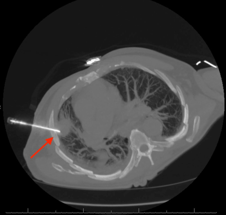
Here, we discuss the case of a 72-year-old male with a known history of COPD who presented with one month of binocular diplopia and headache. The initial clinical investigation discovered destructive intraosseous lesions within the sellar and para-sellar (SPS) regions, suggesting primary versus metastatic intracranial lesions. Further examination revealed a mass in the right lung, with subsequent biopsy confirming squamous cell carcinoma (SCC) of the lung as the primary site of malignancy. The SPS regions of the basicranium, while well-documented to be associated with various primary neoplasms, rarely serve as sites of metastasis. Throughout this article, we will review the pathophysiology of squamous cell lung cancer, current understandings of SPS metastasis, and considerations of metastatic lung SCC management.
Keywords: binocular diplopia; metastatic squamous cell carcinoma; sellar metastasis; squamous cell carcinoma of the lung; squamous cell lung carcinoma.
Copyright © 2022, Sugg et al.
Conflict of interest statement
The authors have declared that no competing interests exist.
Figures


Similar articles
-
An unusual initial presentation of hepatocellular carcinoma as a sellar mass.J Nat Sci Biol Med. 2015 Jul-Dec;6(2):471-4. doi: 10.4103/0976-9668.160045. J Nat Sci Biol Med. 2015. PMID: 26283857 Free PMC article.
-
Unusual case of cavitary lung metastasis from squamous cell carcinoma of the uterine cervix.Pan Afr Med J. 2013;14:37. doi: 10.11604/pamj.2013.14.37.1420. Epub 2013 Jan 27. Pan Afr Med J. 2013. PMID: 23560120 Free PMC article.
-
Primary lung squamous cell carcinoma and its association with gastric metastasis: A case report and literature review.Thorac Cancer. 2020 Jun;11(6):1708-1711. doi: 10.1111/1759-7714.13410. Epub 2020 Mar 25. Thorac Cancer. 2020. PMID: 32212371 Free PMC article. Review.
-
Folliculotropic Cutaneous Metastases and Lymphangitis Carcinomatosa: When Cutaneous Metastases of Breast Carcinoma Are Mistaken for Cutaneous Infections.Acta Dermatovenerol Croat. 2016 Jun;24(2):154-7. Acta Dermatovenerol Croat. 2016. PMID: 27477179
-
Bronchogenic squamous cell carcinoma with soft-tissue metastasis to the hand: an unusual case presentation and review of the literature.Am J Orthop (Belle Mead NJ). 2014 Dec;43(12):E324-7. Am J Orthop (Belle Mead NJ). 2014. PMID: 25490021 Review.
References
Publication types
LinkOut - more resources
Full Text Sources
Research Materials