

Temporomandibular Joint Space Dimensions among Saudi Patients with Temporomandibular Disorders: MRI-Based Retrospective Study
- PMID: 35989869
- PMCID: PMC9363928
- DOI: 10.1155/2022/5846255
Temporomandibular Joint Space Dimensions among Saudi Patients with Temporomandibular Disorders: MRI-Based Retrospective Study
Abstract
Introduction: The temporomandibular joint is a complex synovial joint in the body. It is the area in which the mandible articulates with the cranium. The temporomandibular joint space is located between the articular eminence and the glenoid fossa of the temporal bone at the base of the skull and the condylar process of the mandible. This interarticular space is divided into superior joint space (1.2 ml) and inferior joint space (0.9 ml) by the articular disc. The purpose of this study is to detect and evaluate the variations in the temporomandibular joint space among patients having temporomandibular joint disorders.
Materials and methods: In this retrospective study, 60 magnetic resonance imaging scans were evaluated at King Faisal Specialist Hospital in Riyadh, Saudi Arabia, between the years 2006 and 2016. Measurements were done in sagittal view in three areas: anterior, central, and posterior areas. However, coronal view readings were recorded in two different areas: medial and lateral joint spaces. All measurements were recorded at the highest point of the condyle that is perpendicular to the opposing bone. The SPSS program was used for statistical analysis.
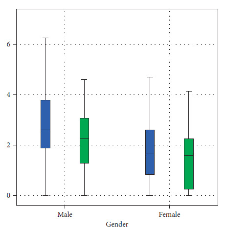
Results: The central joint space values were higher than the anterior and posterior joint spaces in both coronal and sagittal views. We also found that joint spaces among male patients were higher than female patients (right side P=0.015 and left side P=0.006). It is worth mentioning that the number of temporomandibular joint disorder female patients was more than the number of male temporomandibular joint disorder patients (52 females versus 24 males). Additionally, patients who were older than 55 years old had wider joint spaces than patients who were younger than 25 years old.
Conclusion: The central joint space value was the highest among the other joint spaces on both views of magnetic resonance imaging, and the values of joint spaces among males were larger than those of females on sagittal magnetic resonance imaging. Patients with elderly temporomandibular joint disorders showed larger joint spaces than young patients. This study spotlights the importance of magnetic resonance imaging evaluation in temporomandibular joint disorder patients for a better understanding of the clinical evolution of temporomandibular disorders.
Copyright © 2022 Nasser Raqe Alqhtani et al.
Conflict of interest statement
The authors declare that they have no conflicts of interest.
Figures


References
-
- Okeson J. P. Management of Temporomandibular Disorders and Occlusion-E-Book . Amsterdam, Netherlands: Elsevier Health Sciences; 2019.
-
- Okeson J. P. Fundamentals of Occlusion and Temporomandibular Disorders . 4th. São Paulo, Brazil: Art Med; 2020.
-
- Latarjet M., Ruiz-Liard A. Anatomía Humana . 4th. Madrid, Spain: Panamericana; 2007.
MeSH terms
LinkOut - more resources
Full Text Sources
Medical