. 2022 Jul;8(7):e202100349.
doi: 10.1002/cnma.202100349. Epub 2021 Nov 1.

Electrospun Nanofibers for Wound Management

Affiliations

Electrospun Nanofibers for Wound Management

Johnson V John et al. ChemNanoMat. 2022 Jul.

Abstract

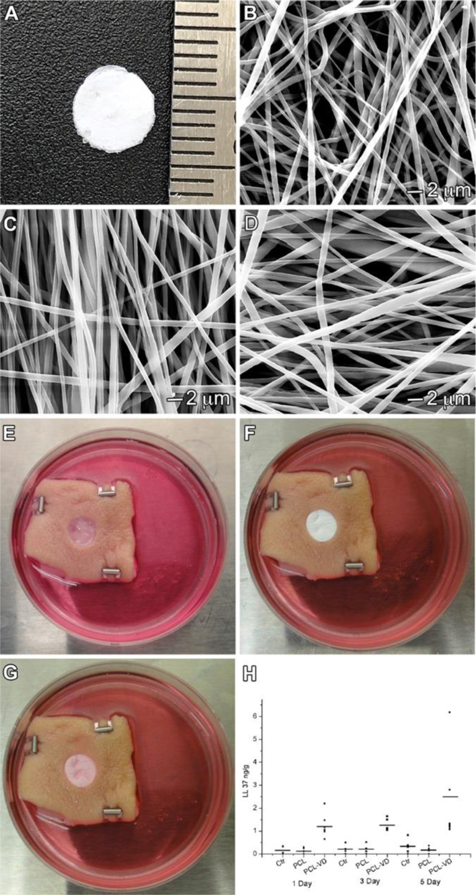
Electrospun nanofibers show great potential in biomedical applications. This mini review article traces the recent advances in electrospun nanofibers for wound management via various approaches. Initially, we provide a short note on the four phases of wound healing, including hemostasis, inflammation, proliferation, and remodeling. Then, we state how the nanofiber dressings can stop bleeding and reduce the pain. Following that, we discuss the delivery of therapeutics and cells using different types of nanofibers for enhancing cell migration, angiogenesis, and re-epithelialization, resulting in the promotion of wound healing. Finally, we present the conclusions and future perspectives regarding the use of electrospun nanofibers for wound management.

Keywords: Antibacterial; Electrospun nanofibers; Hemostasis; Pain relief; Wound healing.


Conflict of interest statement

Conflict of Interest The authors declare no conflict of interest.

Figures

Figure 1.
Figure 1.
Stages of wound healing and their major cellular components. (A) Wound healing begins with hemostasis, where a platelet plug prevents blood loss, and a preliminary fibrin matrix is formed. (B) Inflammation then ensures to remove debris and prevent infection and starts with an influx of neutrophils, which is promoted by histamine release from mast cells. Monocytes arrive later and differentiate into tissue macrophages to clear remaining cell debris and neutrophils. (C) During the proliferative phase, keratinocytes migrate to close the wound gap, blood vessels reform through angiogenesis, and fibroblasts replace the initial fibrin clot with granulation tissue. (D) Finally, the deposited matrix is remodeled further by fibroblasts, blood vessels regress, and myofibroblasts cause overall wound contraction.
Figure 2.
Figure 2.
Hemostatic plug formation in wound healing. Platelet activation cascade leads to hemostatic plug formation, and the coagulation cascade contributes to the stabilization of the thrombus. Adapted with permission from Ref. [12]. Copyright 2015 Elsevier.
Figure 3.
Figure 3.
Endogenous antimicrobial peptide production using 1,25(OH)2D3-loaded PCL nanofiber membranes. (A) Photograph shows a 1,25(OH)2D3-loaded PCL fiber membrane with a diameter of 5 mm. (B-D) SEM images of PCL fibers, 1,25(OH)2D3-loaded PCL fibers and 1,25(OH)2D3-loaded PCL/pluronic F127 fibers. (E) An artificial wound (epidermal and partial dermal layer 1-mm thick) with a diameter of 8 mm was created in each skin explant (Ctr: without any treatment). (F) Dressings containing either 1,25(OH)2D3-loaded PCL fibers or vehicle (PCL fibers) were placed in the wound. (G) The appearance of the skin tissue and nanofiber dressing after five days of culture. (H) Quantification of hCAP18/LL-37 production by ELISA after treatment for 1, 3, and 5 days. Adapted with permission from Ref. [52]. Copyright 2015 Future Medicine Group.
Figure 4.
Figure 4.
Schematic illustrating therapeutics-loaded nanofibers for wound healing. (A) Different approaches used for loading therapeutic molecules to nanofibers. (B) Schematic representation of the incorporated of dimethyloxalylglycine-loaded mesoporous nanoparticles to electrospun nanofibers for wound healing. Adapted with permission from Ref. [93]. Copyright 2018 Elsevier.
Figure 5.
Figure 5.
Growth factors induced wound healing. (A) Schematic illustrating the fabrication and use of nanoparticle-embedded electrospun nanofibers loaded with two growth factors. (B) Characterization of nanoparticle-embedded electrospun nanofibers. (a) SEM images of nanofiber scaffolds: 2:1 CS/PEO-NPs, (b) 1:1 CS/PEO-NPs. (c) Fluorescent image merges monochrome image of ICG-loaded NPs in CS/PEO fibers, as indicated by arrows. (C) Histological evaluation of wounds treated by CS/PEO-NP meshes. Adapted with permission from Ref. [102]. Copyright 2013 Elsevier.
Figure 6.
Figure 6.
Orientation of nanofiber in wound healing. (A) (a-c) SEM images of the different orientations of nanofiber. (B) (a-f) Morphology of fibroblasts cultured for 24 h on nanofibrous scaffolds. Fluorescent staining of F-actin (green) with phalloidin-FITC. Cell nuclei are stained blue with DAPI. (C) H & E staining of wound sections at various time points. Wound areas are traced with a dashed white line. Adapted with permission from Ref. [106]. Copyright 2018 Royal Society of Chemistry.
Figure 7.
Figure 7.
3D nanofibrous scaffolds for wound healing. A (a-d) Top and side view of 1-mm thick radially aligned scaffold and (e-h) Top and side view of 1-mm thick vertically aligned scaffold. Double-headed arrows indicate the alignment of nanofibers. B (a) Schematic illustrating the application of 3D scaffolds consisting of radially aligned nanofibers in stages 1 & 2 DFU healing, which aim to accelerate the re-epithelialization of superficial wounds, (b) Schematic illustrating the application of 3D scaffolds consisting of vertically aligned nanofibers in stages 3 & 4 DFU healing, which aim to promote the formation of granulation tissues of deep wounds, (c) The potential mechanism of 3D scaffolds consisting of radially aligned nanofibers for diabetic wound healing, including enhancing angiogenesis, granulation tissue formation, ECM deposition, and re-epithelialization, and (d) The potential mechanism of 3D scaffolds consisting of vertically aligned nanofibers for diabetic wound healing, including enhancing angiogenesis, granulation tissue formation, and ECM deposition. Adapted with permission from Ref. [108]. Copyright 2020 Elsevier.
Figure 8.
Figure 8.
Fibroblast culture on nanofiber membranes with different organizations. NIH 3T3 fibroblasts were seeded on aligned (A), random (B), microwell (C), and flat well (D) nanofiber membranes and incubated for 3, 7, 10, 14, and 21 days. Insets illustrated cell suspension droplets on the surface of nanofiber membranes. The distance between the two adjacent microwells was 3 mm. Fluorescence microscopy images showed living cells cultured on nanofiber membranes which were stained with fluorescein diacetate in green. All scale bars are 1 mm. Adapted with permission from Ref. [117]. Copyright 2014 Elsevier.
Figure 9.
Figure 9.
Nanofiber skin grafts for wound healing. (A) Transplanted microskins indicated by small black arrows in sandwich-type nanofiber scaffolds were transplanted satisfactorily on wounds with a uniform distribution at day seven post-surgery. (B) Re-epithelialization derived from microskins occurred along the wound bed on day 14 after surgery. (C) The wound was completely closed by re-epithelialization derived from microskins indicated by black arrows on day 21 after surgery. (D) Magnified view of the region d in (A), transplanted microskins contained both epidermal and dermal layers indicated by white dashed lines and white arrowheads, respectively, confined by the nanofiber microwell indicated by black dashed lines. (E) Magnified view of the region e in (D), showing small blood vessels indicated by white arrowheads, large collagen bundles, and a few fibroblasts in the dermal layer of the microskin. (F) Magnified view of the region f in (B), showing stratified epithelial cells derived from microskins crept along the surface of wound bed toward the adjacent microskin indicated by white dash lines. Simultaneously, the dermal layer of microskins began integrating with the wound bed indicated by white arrowheads. (G) Magnified view of the region g in (C), showing epidermal cells migrated from the two adjacent micro skin resurfaced the wound indicated by white dash lines Adapted with permission from Ref. [117]. Copyright 2014 Elsevier.

References

    1. Broughton G, Janis JE, Attinger CE, Plast Reconstr. Surg 2006, 117, 12S–34S. - PubMed
    1. Ziegler TR, Pierce GF, Herndon DN, Growth Factors and Wound Healing New York: Springer, 2012, 1.
    1. Attinger CE, Janis JE, Steinberg J, Schwartz J, Al-Attar A, Couch K, Plast. Reconstr. Surg 2006, 117, 72S–109S. - PubMed
    1. Bandyk DF, Semin. Vasc. Surg 2018, 31, 43–48. - PubMed
    1. Ahmad J, Diabetes Metab. Syndr 2016, 10, 48–60. - PubMed

LinkOut - more resources