

Arrhythmogenic Mitral Valve Prolapse
- PMID: 35990107
- PMCID: PMC9376835
- DOI: 10.15420/aer.2021.28
Arrhythmogenic Mitral Valve Prolapse
Abstract
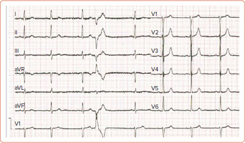
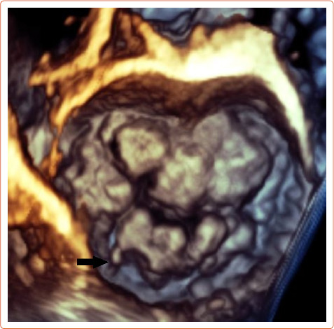
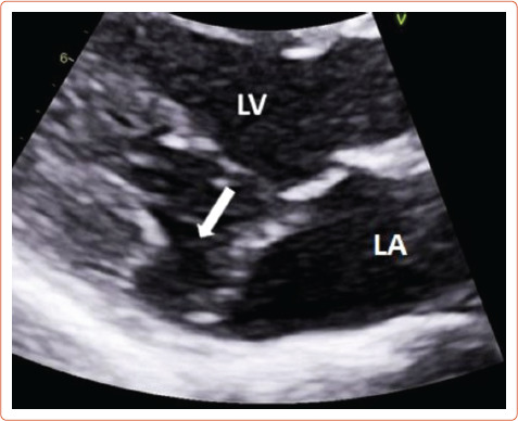
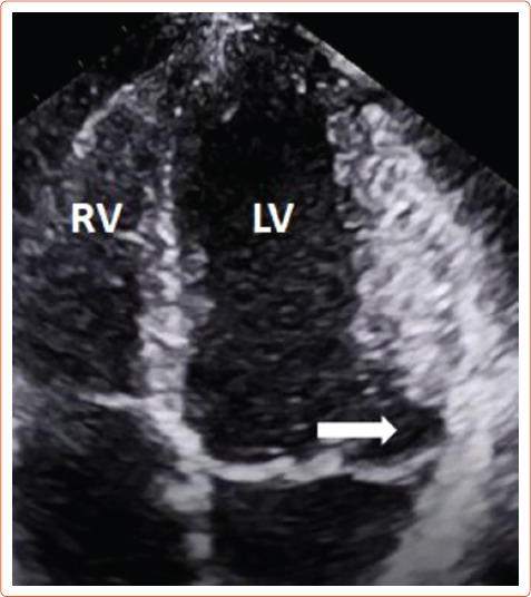
Mitral valve prolapse (MVP) is a common condition present in 1-3% of the population. There has been evidence that a subset of MVP patients is at higher risk of sudden cardiac death. The arrhythmogenic mechanism is related to fibrotic changes in the papillary muscles caused by the prolapsing valve. ECG features include ST-segment depression, T wave inversion or biphasic T waves in inferior leads, and premature ventricular contractions arising from the papillary muscles and the fascicular system. Echocardiography can identify MVP and mitral annular disjunction, a feature that has significant negative prognostic value in MVP. Cardiac MRI is indicated for identifying fibrosis. Patients with high-risk features should be referred for further evaluation. Catheter ablation and mitral valve repair might reduce the risk of malignant arrhythmia. MVP patients with high-risk features and clinically documented ventricular arrhythmia may also be considered for an ICD.
Keywords: Arrhythmia; echocardiography; mitral annular disjunction; mitral valve prolapse; papillary muscle fibrosis; premature ventricular contractions; sudden cardiac death.
Copyright © 2022, Radcliffe Cardiology.
Conflict of interest statement
Disclosure: DGK is editor-in-chief of Arrhythmia & Electrophysiology Review; this did not influence peer review. All other authors have no conflicts of interest to declare.
Figures

Similar articles
-
Papillary muscle ventricular ectopy ablation in a malignant mitral valve prolapse: Ablation in Malignant Mitral Valve Prolapse: Ablation in Malignant Mitral Valve Prolapse.Echocardiography. 2023 Mar;40(3):271-275. doi: 10.1111/echo.15529. Epub 2023 Jan 31. Echocardiography. 2023. PMID: 36722012
-
Mitral Valve Prolapse and Sudden Cardiac Death in Athletes at High Risk.Curr Cardiol Rev. 2023;19(3):e201222212066. doi: 10.2174/1573403X19666221220163431. Curr Cardiol Rev. 2023. PMID: 36545732 Free PMC article.
-
Transthoracic echocardiography for arrhythmic mitral valve prolapse: Phenotypic characterization as first step.Echocardiography. 2022 Sep;39(9):1158-1170. doi: 10.1111/echo.15439. Epub 2022 Aug 27. Echocardiography. 2022. PMID: 36029124 Review.
-
Abnormal Mechanics Relate to Myocardial Fibrosis and Ventricular Arrhythmias in Patients With Mitral Valve Prolapse.Circ Cardiovasc Imaging. 2023 Apr;16(4):e014963. doi: 10.1161/CIRCIMAGING.122.014963. Epub 2023 Apr 18. Circ Cardiovasc Imaging. 2023. PMID: 37071717 Free PMC article.
-
Mitral Valve Prolapse and Mitral Annular Disjunction Arrhythmic Syndromes: Diagnosis, Risk Stratification and Management.Rev Cardiovasc Med. 2022 Sep 5;23(9):295. doi: 10.31083/j.rcm2309295. eCollection 2022 Sep. Rev Cardiovasc Med. 2022. PMID: 39077697 Free PMC article. Review.
Cited by
-
Arrhythmogenic Mitral Valve Prolapse: Can We Risk Stratify and Prevent Sudden Cardiac Death?Arrhythm Electrophysiol Rev. 2024 Jul 23;13:e11. doi: 10.15420/aer.2023.26. eCollection 2024. Arrhythm Electrophysiol Rev. 2024. PMID: 39145277 Free PMC article. Review.
-
Brazilian Guideline for Exercise Testing in Children and Adolescents - 2024.Arq Bras Cardiol. 2024 Sep 16;121(8):e20240525. doi: 10.36660/abc.20240525. Arq Bras Cardiol. 2024. PMID: 39292116 Free PMC article. English, Portuguese.
-
Broad Electrocardiogram Syndromes Spectrum: From Common Emergencies to Particular Electrical Heart Disorders-Part II.Diagnostics (Basel). 2025 Jun 19;15(12):1568. doi: 10.3390/diagnostics15121568. Diagnostics (Basel). 2025. PMID: 40564888 Free PMC article. Review.
-
Biology of mitral valve prolapse: from general mechanisms to advanced molecular patterns-a narrative review.Front Cardiovasc Med. 2023 Jun 2;10:1128195. doi: 10.3389/fcvm.2023.1128195. eCollection 2023. Front Cardiovasc Med. 2023. PMID: 37332582 Free PMC article. Review.
-
Mitral Valve Prolapse With Mitral Annular Disjunction Causing Ventricular Tachycardia During a Stress Test: A Case Report.Cureus. 2024 Jul 7;16(7):e64020. doi: 10.7759/cureus.64020. eCollection 2024 Jul. Cureus. 2024. PMID: 39109096 Free PMC article.
References
-
- Otto CM, Nishimura RA, Bonow RO et al. 2020 ACC/AHA guideline for the management of patients with valvular heart disease: executive summary: a report of the American College of Cardiology/American Heart Association Joint Committee on Clinical Practice guidelines. Circulation. 2021;143:e35–71. doi: 10.1161/CIR.0000000000000932. - DOI - PubMed
Publication types
LinkOut - more resources
Full Text Sources
Miscellaneous
