

Predicting Hematoma Expansion after Spontaneous Intracranial Hemorrhage Through a Radiomics Based Model
- PMID: 35990197
- PMCID: PMC9390077
- DOI: 10.1117/12.2611847
Predicting Hematoma Expansion after Spontaneous Intracranial Hemorrhage Through a Radiomics Based Model
Abstract
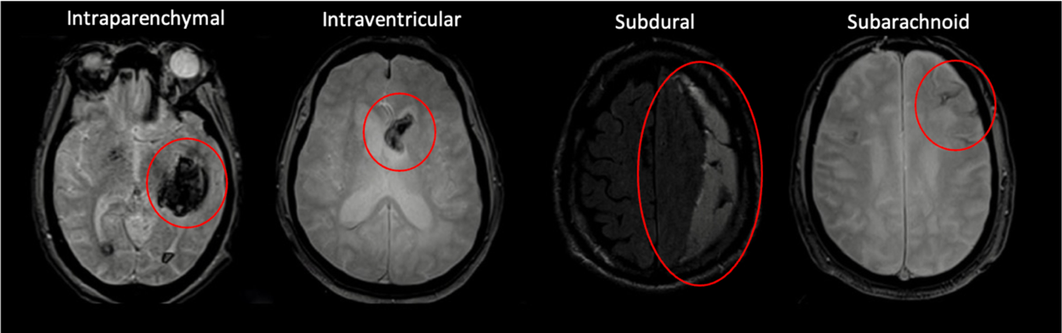
Purpose: Intracranial hemorrhage (ICH) is characterized as bleeding into the brain tissue, intracranial space, and ventricles and is the second most disabling form of stroke. Hematoma expansion (HE) following ICH has been correlated with significant neurological decline and death. For early detection of patients at risk, deep learning prediction models were developed to predict whether hematoma due to ICH will expand. This study aimed to explore the feasibility of HE prediction using a radiomic approach to help clinicians better stratify HE patients and tailor intensive therapies timely and effectively.
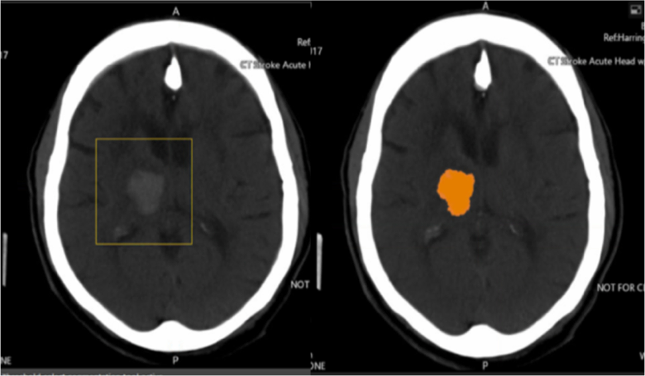
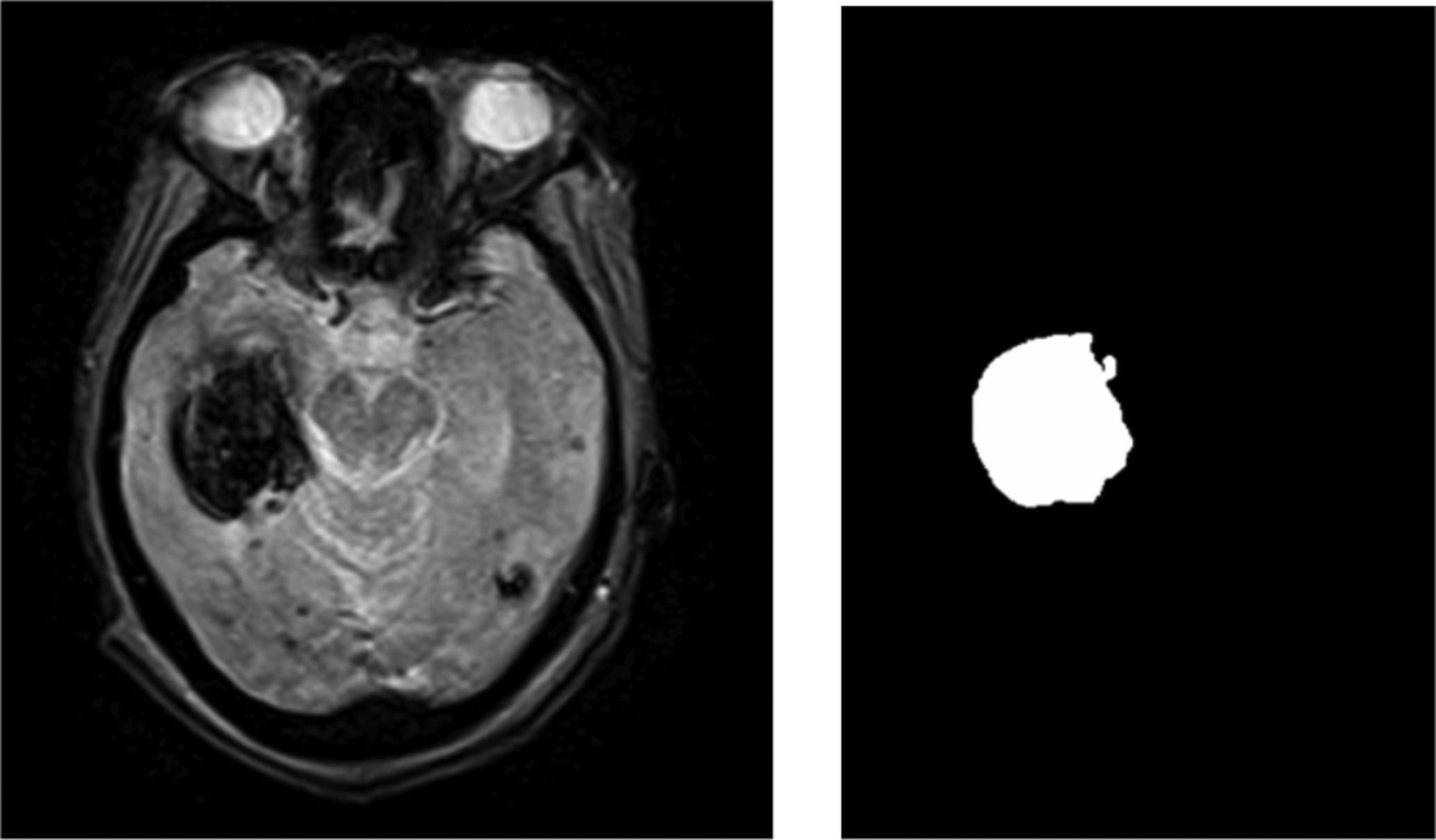
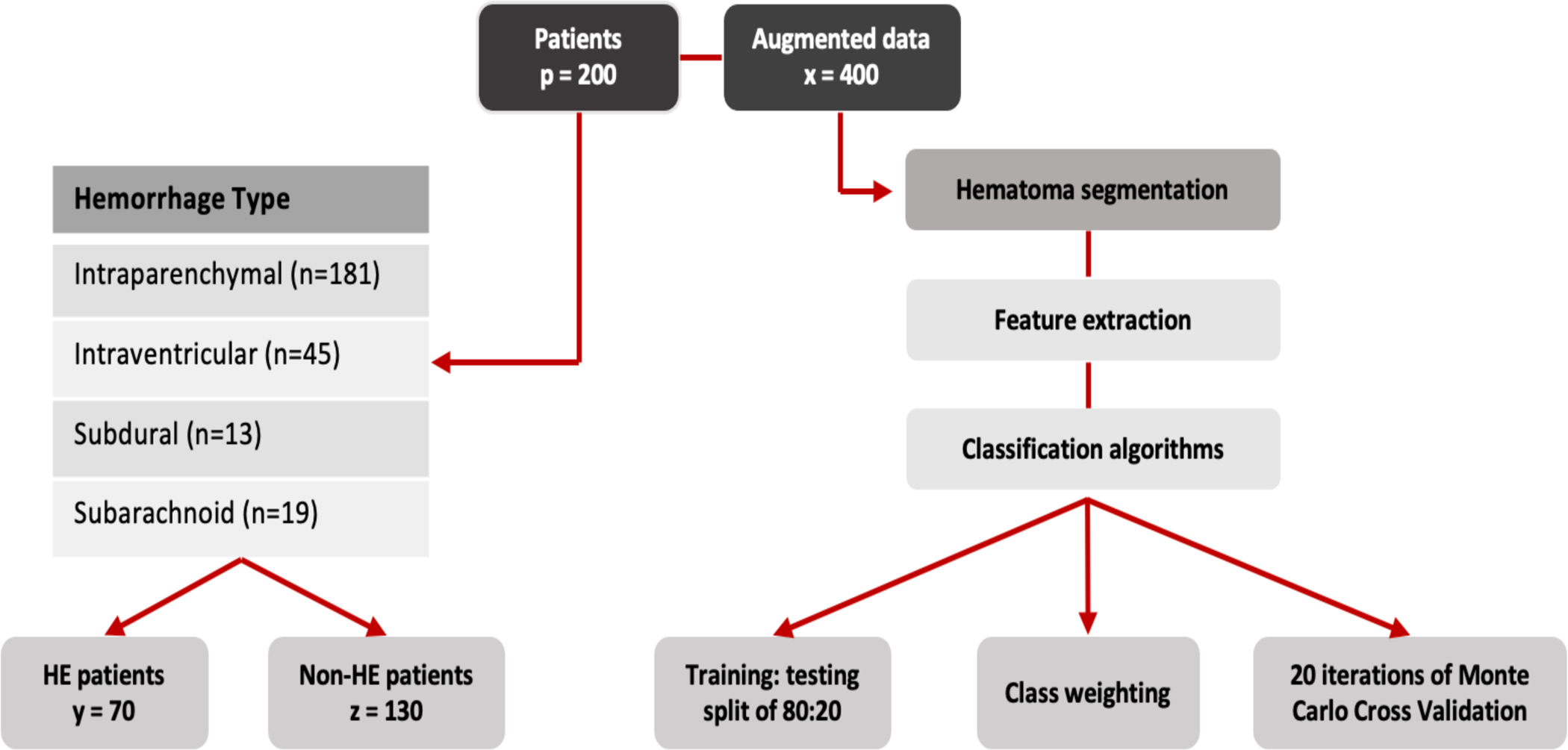
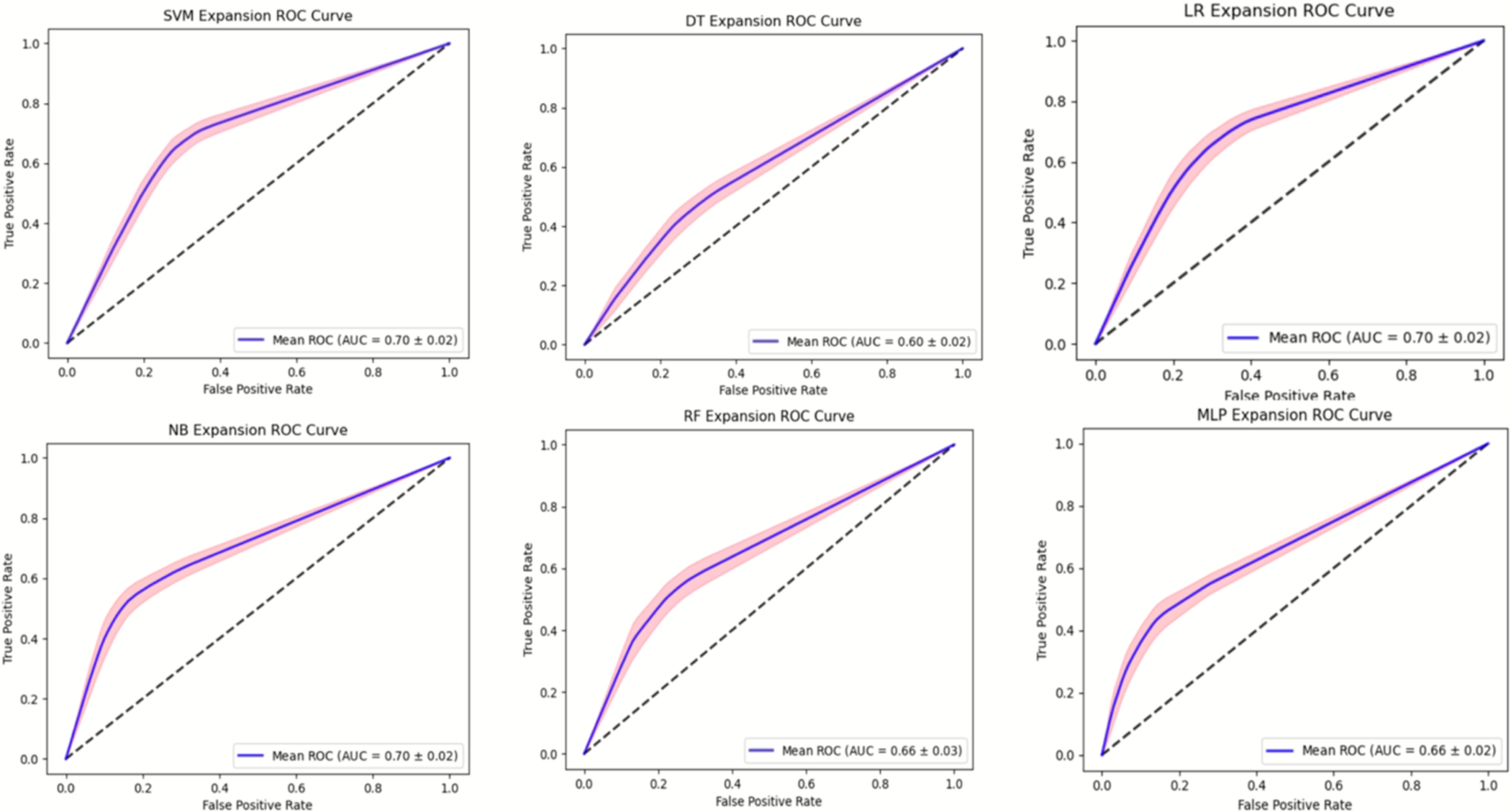
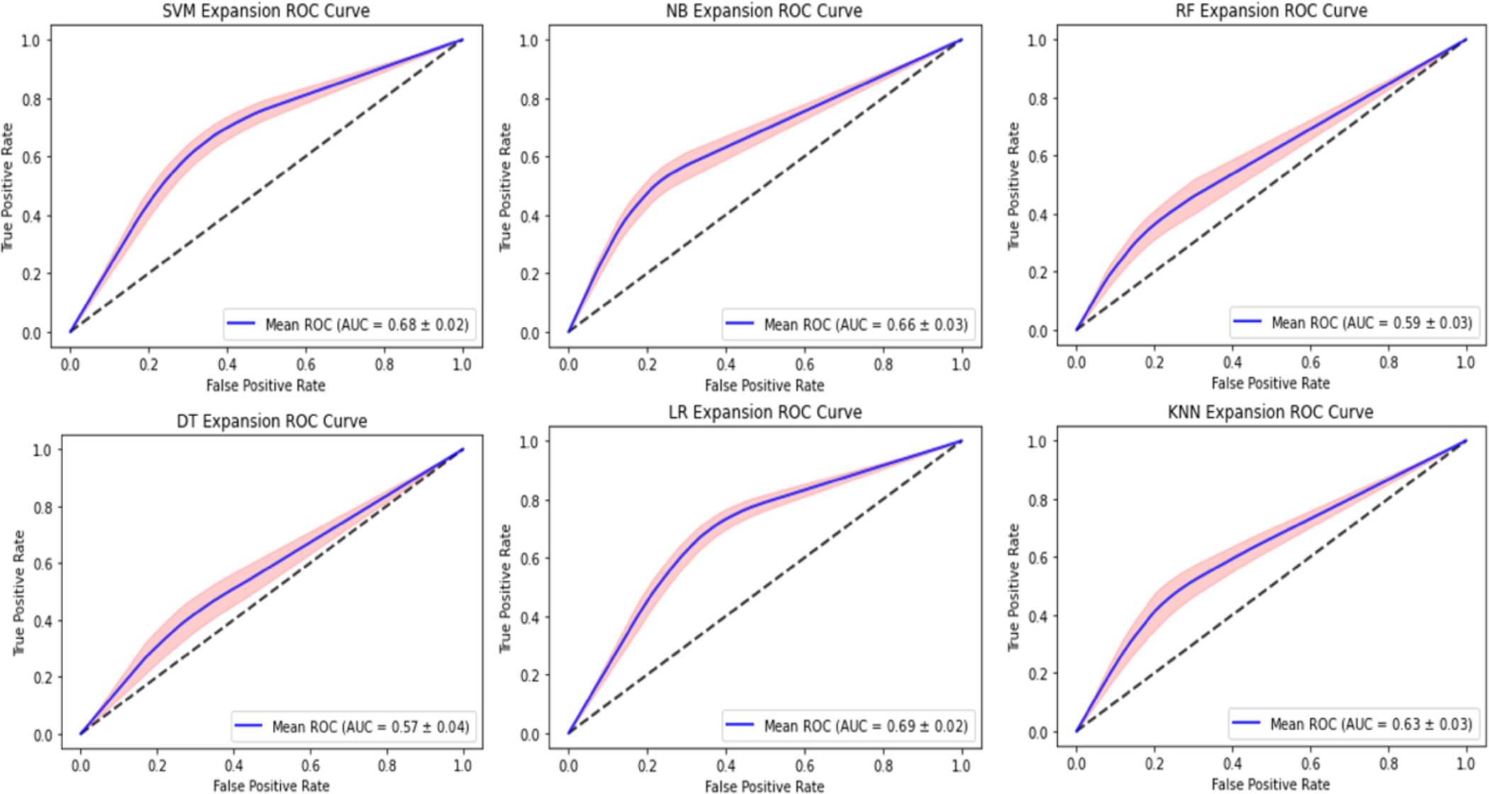
Materials and methods: Two hundred ICH patients with known hematoma evolution, were enrolled in this study. An open-source python package was utilized for the extraction of radiomic features from both non-contrast computed tomography (NCCT) and magnetic resonance imaging (MRI) scans through characterization algorithms. A total of 99 radiomic features were extracted and different features were selected for network inputs for the NCCT and MR models. Seven supervised classifiers: Support Vector Machine, Naïve Bayes, Decision Tree, Random Forest, Logistic Regression, K-Nearest Neighbor and Multilayer Perceptron were used to build the models. A training:testing split of 80:20 and 20 iterations of Monte Carlo cross validation were performed to prevent overfitting and assess the variability of the networks, respectively. The models were fed training datasets from which they learned to classify the data based on pre-determined radiomic categories.
Results: The highest sensitivity among the NCCT classifier models was seen with the support vector machine (SVM) and logistic regression (LR) of 72 ± 0.3% and 73 ± 0.5%, respectively. The MRI classifier models had the highest sensitivity of 68 ± 0.5% and 72 ± 0.5% for the SVM and LR models, respectively.
Conclusions: This study indicates that the NCCT radiomics model is a better predictor of HE and that SVM and LR classifiers are better predictors of HE due to their more cautious approach indicated by a higher sensitivity metric.
Keywords: Artificial Intelligence; Brain; Hematoma Expansion; Hemorrhagic Stroke; Non-contrast Computed Tomography.
Figures
Similar articles
-
Clinical Features, Non-Contrast CT Radiomic and Radiological Signs in Models for the Prediction of Hematoma Expansion in Intracerebral Hemorrhage.Can Assoc Radiol J. 2023 Nov;74(4):713-722. doi: 10.1177/08465371231168383. Epub 2023 Apr 18. Can Assoc Radiol J. 2023. PMID: 37070854
-
Comparison of Radiomic Models Based on Different Machine Learning Methods for Predicting Intracerebral Hemorrhage Expansion.Clin Neuroradiol. 2022 Mar;32(1):215-223. doi: 10.1007/s00062-021-01040-2. Epub 2021 Jun 22. Clin Neuroradiol. 2022. PMID: 34156513
-
Quantitative imaging for predicting hematoma expansion in intracerebral hemorrhage: A multimodel comparison.J Stroke Cerebrovasc Dis. 2024 Jul;33(7):107731. doi: 10.1016/j.jstrokecerebrovasdis.2024.107731. Epub 2024 Apr 23. J Stroke Cerebrovasc Dis. 2024. PMID: 38657831
-
Machine learning for predicting hematoma expansion in spontaneous intracerebral hemorrhage: a systematic review and meta-analysis.Neuroradiology. 2024 Sep;66(9):1603-1616. doi: 10.1007/s00234-024-03399-8. Epub 2024 Jun 12. Neuroradiology. 2024. PMID: 38862772
-
Efficacy of non-enhanced computer tomography-based radiomics for predicting hematoma expansion: A meta-analysis.Front Oncol. 2023 Jan 10;12:973104. doi: 10.3389/fonc.2022.973104. eCollection 2022. Front Oncol. 2023. PMID: 36703784 Free PMC article.
Cited by
-
Need for Transparency and Clinical Interpretability in Hemorrhagic Stroke Artificial Intelligence Research: Promoting Effective Clinical Application.Yonsei Med J. 2024 Oct;65(10):611-618. doi: 10.3349/ymj.2024.0007. Yonsei Med J. 2024. PMID: 39313452 Free PMC article.
-
The clinical potential of radiomics to predict hematoma expansion in spontaneous intracerebral hemorrhage: a narrative review.Front Neurol. 2024 Jul 19;15:1427555. doi: 10.3389/fneur.2024.1427555. eCollection 2024. Front Neurol. 2024. PMID: 39099779 Free PMC article. Review.
-
Research advances in predicting the expansion of hypertensive intracerebral hemorrhage based on CT images: an overview.PeerJ. 2024 Jun 7;12:e17556. doi: 10.7717/peerj.17556. eCollection 2024. PeerJ. 2024. PMID: 38860211 Free PMC article. Review.
References
-
- Zheng Y, Hu Q, Manaenko A, Zhang Y, Peng Y, Xu L, Tang J, Tang J, & Zhang JH (2015). 17β-Estradiol attenuates hematoma expansion through estrogen receptor α/silent information regulator 1/nuclear factor-kappa b pathway in hyperglycemic intracerebral hemorrhage mice. Stroke, 46(2), 485–491. 10.1161/STROKEAHA.114.006372 - DOI - PMC - PubMed
-
- Griethuysen JJM, Fedorov A, Parmar C, Hosny A, Aucoin N, Narayan V, Beets-Tan RGH, Fillon-Robin JC, Pieper S, Aerts HJWL (2017). Computational Radiomics System to Decode the Radiographic Phenotype. Cancer Research, 77(21), e104–e107. `10.1158/0008-5472.CAN-17-0339 <10.1158/0008-5472.CAN-17-0339>`_ - DOI - DOI - PMC - PubMed
Grants and funding
LinkOut - more resources
Full Text Sources