Case Reports
. 2022 Aug 13:15:1639-1642.
doi: 10.2147/CCID.S378253. eCollection 2022.

Primary Localized Cutaneous Nodular Amyloidosis Presenting as Milia: An Unusual Clinical Manifestation

Affiliations
Case Reports

Primary Localized Cutaneous Nodular Amyloidosis Presenting as Milia: An Unusual Clinical Manifestation

Yanqing Wang et al. Clin Cosmet Investig Dermatol. .

Abstract

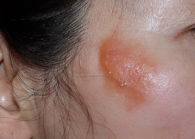
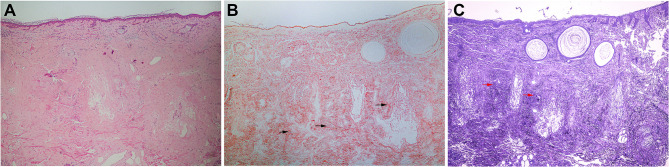
Primary localized cutaneous nodular amyloidosis (PLCNA) is rare and clinically noncharacteristic, presenting mostly as plaque-like lesions. We report a case of a progressively larger erythematous plaque following a contusion of the skin on the right zygomatic area, which was strangely covered with recurrent scattered 2 mm whiteish blisters to the extent that it was misdiagnosed as a herpesvirus infection several times over a decade. Pathology and special staining diagnosed nodular amyloidosis with milia.

Keywords: local trauma; milia; plaque; primary localized cutaneous nodular amyloidosis.


Conflict of interest statement

The authors report no conflicts of interest related to this work.

Figures

Figure 1
Figure 1
Orange‒yellow plaque with overlying scattered 2 mm whiteish blisters on the right zygomatic area.
Figure 2
Figure 2
Three pathological images of nodular amyloidosis. (A) Deposition of acellular amorphous eosinophilic materials over the entire dermis and subcutis (H&E, ×200). (B) The amyloid deposits were positive for Congo red staining, and keratinous cysts were found in the superficial dermis. The arrows indicate the sites of amyloid deposits.(Congo red staining, ×200) (C) The amyloid deposits were positive for crystal violet staining, and keratinous cysts were found in the superficial dermis. The arrows indicate the sites of amyloid deposits. (Crystal violet staining, ×200).

Similar articles

Cited by

References

    1. Mena L, Carrasco C, Folch H, de la Parra R, Carreño L. Primary cutaneous nodular amyloidosis associated with the injection of autologous fat. Clin Exp Dermatol. 2021;46:552–554. doi:10.1111/ced.14429 - DOI - PubMed
    1. Vestery JP, Tidman MJ, Mclaren KM. Primary nodular cutaneous amyloidosis – long-term follow-up and treatment. Clin Exp Dermatol. 1994;19:159–162. doi:10.1111/j.1365-2230.1994.tb01148.x - DOI - PubMed
    1. Ung CY, Carr NJ, Ardern-Jones MR. Primary cutaneous nodular amyloidosis associated with psoriasis. Clin Exp Dermatol. 2014;39:608–611. doi:10.1111/ced.12347 - DOI - PubMed
    1. Dickison P, Howard V, Wylie B, Smith SD. Localized axillary milia en plaque: a rare cutaneous case presentation of systemic amyloidosis. Clin Exp Dermatol. 2016;41(7):764–767. doi:10.1111/ced.12914 - DOI - PubMed
    1. Yahya H. Idiopathic multiple eruptive milia: report of a case in a Nigerian woman. Niger J Clin Pract. 2018;21:395–396. doi:10.4103/njcp.njcp_43_17 - DOI - PubMed

Publication types