. 2022 Aug 5:16:916510.
doi: 10.3389/fnana.2022.916510. eCollection 2022.

Expression of Opsins of the Box Jellyfish Tripedalia cystophora Reveals the First Photopigment in Cnidarian Ocelli and Supports the Presence of Photoisomerases

Affiliations

Expression of Opsins of the Box Jellyfish Tripedalia cystophora Reveals the First Photopigment in Cnidarian Ocelli and Supports the Presence of Photoisomerases

Anders Garm et al. Front Neuroanat. .

Erratum in

Abstract

Cubomedusae, or box jellyfish, have a complex visual system comprising 24 eyes of four types. Like other cnidarians, their photoreceptor cells are ciliary in morphology, and a range of different techniques together show that at least two of the eye types-the image-forming upper and lower lens eyes-express opsin as the photopigment. The photoreceptors of these two eye types express the same opsin (Tc LEO), which belongs to the cnidarian-specific clade cnidops. Interestingly, molecular work has found a high number of opsin genes in box jellyfish, especially in the Caribbean species Tripedalia cystophora, most of which are of unknown function. In the current study, we raised antibodies against three out of five opsins identified from transcriptomic data from T. cystophora and used them to map the expression patterns. These expression patterns suggest one opsin as the photopigment in the slit eyes and another as a putative photoisomerase found in photoreceptors of all four eyes types. The last antibody stained nerve-like cells in the tentacles, in connection with nematocytes, and the radial nerve, in connection with the gonads. This is the first time photopigment expression has been localized to the outer segments of the photoreceptors in a cnidarian ocellus (simple eye). The potential presence of a photoisomerase could be another interesting convergence between box jellyfish and vertebrate photoreceptors, but it awaits final experimental proof.

Keywords: box jellyfish; cnidaria; cubozoa; opsin phylogeny; photopigment; phototransduction; vision.


Conflict of interest statement

The authors declare that the research was conducted in the absence of any commercial or financial relationships that could be construed as a potential conflict of interest.

Figures

Figure 1
Figure 1
Box jellyfish T. cystophora. (A) Adult medusa of T. cystophora high lighting the paired gonads (Go) and tentacles (Te). (B) Close-up of a rhopalium showing the four eye types: upper and lower lens eye (ULE, LLE), slit eyes (SE), and pit eyes (PE). Cr, crystal; L, lens. (C) Schematic drawing of a cross section midways in the slit eye [broken line in (B)]. Note the asymmetric groove formed by the pigmented cells housing the outer segments of the ciliary photoreceptors. CR, ciliary rootlet; Nu, nucleus; OS, outer segments; PG, pigment granules. (A) Modified from Bielecki et al. (2013), (C) is modified from Garm et al. (2008).
Figure 2
Figure 2
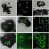
Expression of opsin 1 in T. cystophora. (A) Overlay of stain with antibody against opsin 1 (green) and transmission showing a rhopalium and parts of bell rim of a juvenile medusa. The retina of the slit eye (SE) is brightly stained. (B) The area in (A) only showing the immune stain. Note the complete absence of stain outside the retina of SE. (C) Close-up of SE from (A). The entire retina is rather evenly stained. (D) A juvenile rhopalium seen frontally showing identical staining in the two-slit eyes. (E) Close-up of left SE in (D). Note that the stain is restricted to the photoreceptors outer segments and nor the cell bodies (encircles by broken white line) or the lens-like cells (LLC) are stained. (F) Staining of the retina in an adult SE. LLE, lower lens eye; PE, pit eye; ULE, upper lens eye.
Figure 3
Figure 3
Expression of opsin 2 in T. cystophora. (A) Overlay between transmission and antibody stain of a juvenile rhopalium. Note that the retina of all eyes is stained. (B) The area in (A) only showing the immune stain. The slit eyes (SE) are only weakly stained (arrow). (C) Close-up of pit eye (PE) from (A). The stain is only seen inside the pigment screen (arrowhead) which is the outer segments of the photoreceptors. (D) Close-up of SE from (A). The stain is again only seen in the outer segments (arrowhead) of the photoreceptors inside the pigment screen. (E) Close-up of the upper lens eye (ULE) from (A). Note that the stain in the outer segments is not homogeneous but rather appears as individual lines. (F) Close-up of the lower lens eye (LLE) from (A). (G) Immuno-stain of a juvenile rhopalium clearly showing the staining pattern as lines following the orientation of the segments in the ULE (insert, arrowhead). (H) Stain in an adult PE also showing the expression of opsin 2 as individual lines (arrows). (I) The immuno-stain is also weak in the adult SE (outlines by broken white line) and appears as randomly oriented lines (arrows). (J) The immune stain in the adult ULE is similar to the juvenile ULE [compare with (G)] except for the number of stained lines (arrows) being higher.
Figure 4
Figure 4
Expression of opsin 3 in T. cystophora. (A) Punctuated staining is seen along the midline of the immature gonad (Go, framed by broken white line) in the area where the interradial canal and interradial nerve are also situated. (B) Close-up of framed area in (A) showing the punctuated staining (arrows). (C) The interradial nerve stained with custom-made antibodies raised against Tripedalia RFamide. See Nielsen et al. (2019) for details. (D) Lines of punctuated staining are also found between the autofluorescent nematocyst (Nc) on the tentacles (Te). (E) Close-up of framed area in (C) showing lines of punctuated staining (arrows) between the Nc. Note that the larger nematocysts are not autofluorescent (asterisks).
Figure 5
Figure 5
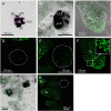
Control staining in T. cystophora. (A,D) Overlay between transmission and staining and staining alone from pre-absorption test with opsin 1 antibodies. Note the complete absence of staining in the rhopalium including the slit eye (Se). Broken white line in (D) encircles the rhopalium. Asterisks indicate autofluorescent nematocysts. (B,E) Overlay between transmission and staining and staining alone from pre-absorption test with opsin 2 antibodies. Note the complete absence of staining in any of the four eye types on the rhopalium [Outlined by broken white line in (E)]. Asterisks indicate autofluorescent nematocysts. (C,F) Overlay between transmission and staining and staining alone from pre-absorption test with opsin 3 antibodies. Note the absence of punctuated staining between the autofluorescent nematocyst on the tentacle (Te) (insert). (G,H) Negative control staining of juvenile medusa. Note the lack of staining in the rhopalium (Rho). Broken white line in (H) encircles the rhopalium. The only fluorescence comes from the autofluorescent nematocysts on the Te. LLE, lower lens eye; PE, pit eye; ULE, upper lens eye.
Figure 6
Figure 6
Staining with opsin 1 antibody in other medusae. (A,D) Overlay between transmission and staining and staining alone of a rhopalium (Rho) of Aurelia aurita. No staining is seen including in the aboral ocellus (Aoc) and the oral ocellus (Ooc). (B,E) Overlay between transmission and staining and staining alone of a rhopalium of Cassiopea xamachana. No staining is seen including ocellus (Oc). (C) Overlay between transmission and staining in the tentacular bulb of Sarsia tubolosa. No staining is seen including ocellus (Oc). (F) A huge number of autofluorescent cells are found on the middle part of the manubrium (Ma, outlined by broken white line) of S. tubolosa. Cr, crystal; EsD, endosymbiotic dinoflagellates; Te, tentacle.
Figure 7
Figure 7
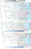
Maximum likelihood phylogenetic analysis of known opsins of the box jellyfish Tripedalia cystophora. We combined 21 protein sequences of opsin of T. cystophora (red) with other known cnidops, including those of Alatina alata (orange), and xenopsins (outgroup in gray), then added putative Morbakka opsin (orange)—for multiple alignments using MAFFT. We then selected the best-fit model in IQ-TREE for likelihood and UFBoot phylogenetic analyses. Illustrated is the bootstrap consensus with numbers at nodes showing bootstrap proportions. Two large clades are collapsed, one containing Anthozoa cnidops, and the other containing mainly Hydrozoa cnidops (see Supplementary Material for full phylogeny). Cubozoan opsins fall into three clades, somewhat consistent with previous analyses, which we label here as Group 1a, Group 1b, and Group 2 (Liegertová et al., 2015). Our antibody experiments labeled Tripedalia cystophora slit eye opsins—Tc SEO (Antibody 1), labeled Tripedalia cystophora general eye opsins—Tc GEO (Antibody 2), and labeled Tripedalia cystophora gonads and cnidocyte opsins—Tc GCO (Antibody 3). Note that both Tc LEO and Tc GCO are represented. These originate from two different studies and putatively represent two different alleles of the same opsin gene.

Similar articles

Cited by

References

    1. Artigas Q. O., Lapebie P., Leclere L., Takeda N., Deguchi R., Jekely G., et al. . (2018). A gonad-expressed opsin mediates light-induced spawning in the jellyfish Clytia. eLife 7, e29555. 10.7554/eLife.29555 - DOI - PMC - PubMed
    1. Bielecki J., Nachman G., Garm A. (2013). Swimpacemaker response to bath applied neurotransmitters in the cubozoan Tripedalia cystophora. J. Comp. Physiol. A 199, 785–795. 10.1007/s00359-013-0839-1 - DOI - PubMed
    1. Bielecki J., Zaharoff A., Leung N., Garm A., Oakley T. H. (2014). The cubozoan visual system utilizes several opsins. PLoS ONE 9, 1–9. 10.1371/journal.pone.0098870 - DOI - PMC - PubMed
    1. Döring C. C., Kumer S., Tumu S. C., Kourtesis I., Hausen H. (2020). The visual pigment xenopsin is widespread in protostome eyes and impacts the view on eye evolution. eLife 9:e55193. 10.7554/eLife.55193 - DOI - PMC - PubMed
    1. Ekström P., Garm A., Pålsson J., Vihtlec T., Nilsson D. E. (2008). Immunohistochemical evidence for several photosystems in box jellyfish using opsin-antibodies. Cell Tissue Res. 333, 115–124. 10.1007/s00441-008-0614-8 - DOI - PubMed

LinkOut - more resources