Sacubitril/Valsartan Improves Progression of Early Diabetic Nephropathy in Rats Through Inhibition of NLRP3 Inflammasome Pathway
- PMID: 35992034
- PMCID: PMC9386175
- DOI: 10.2147/DMSO.S366518
Sacubitril/Valsartan Improves Progression of Early Diabetic Nephropathy in Rats Through Inhibition of NLRP3 Inflammasome Pathway
Abstract
Purpose: Diabetic nephropathy (DN), a global disease, is the leading cause of end-stage renal disease. There is a lack of specific treatment for this disease, and early intervention in disease progression is essential. In this paper, we used a rat model of early diabetic nephropathy to explore the therapeutic mechanism of sacubitril/valsartan in rats with early diabetic nephropathy.
Materials and methods: Rats were grouped into 1 normal group; 2. Model group (DN group): STZ (45 mg/kg/d) induced early diabetic nephropathy rats; 3. Sac group: DN rats + Sac group (orally, 60 mg/kg/d) for 6 weeks. After 6 weeks, the levels of serum albumin (ALB), glucose (GLU), creatinine (Cr), urea nitrogen (BUN) and 24-h urinary protein excretion were measured. In renal tissue homogenates, NLRP3 inflammasome, proinflammatory factors IL1-β and TNF-α, oxidative stress MDA and pro-fibrotic cytokine TGF-β1 were performed. Histological analysis of kidneys by hematoxylin and eosin (HE), PAS and Masson trichrome staining.
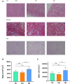
Results: 1. Sacubitril/valsartan (Sac) significantly improved renal hypertrophy, proteinuria and serum albumin levels in rats with early diabetic nephropathy (P < 0.001), and decreased GLU, Scr (P<0.001), and BUN levels (P < 0.01).2. Light microscopy of renal tissues showed glomerular hypertrophy and interstitial inflammatory cell infiltration, and mean glomerular area (MGA) and mean glomerular volume (MGV) were crucially increased in early diabetic nephropathy (P < 0.001), and the Sac group showed reduced renal pathology and improved MGA and MGV (P < 0.001).3. Kidney tissue homogenate levels of NLRP3, Caspase-1, IL1-β, TNF-α, MDA and TGF-β1 were critically, increased in DN rats (P < 0.001), and SOD was significantly decreased. All these indicators above were improved after treatment (P < 0.0001).
Conclusion: Nlrp3-inflammasome promote progression of diabetic nephropathy through inflammation, fibrosis and oxidative stress; sacubitril/valsartan ameliorated early diabetes-induced renal damage by inhibiting NLRP3 pathway activation.
Keywords: Nlrp3-inflammasome; diabetic nephropathy; fibrosis; inflammation; oxidative stress; sacubitril/valsartan.
© 2022 Pan et al.
Conflict of interest statement
The authors have no conflicts of interest to declare.
Figures


Similar articles
-
LCZ696 mitigates diabetic-induced nephropathy through inhibiting oxidative stress, NF-κB mediated inflammation and glomerulosclerosis in rats.PeerJ. 2020 Jun 19;8:e9196. doi: 10.7717/peerj.9196. eCollection 2020. PeerJ. 2020. PMID: 32596035 Free PMC article.
-
Huangkui capsule alleviates renal tubular epithelial-mesenchymal transition in diabetic nephropathy via inhibiting NLRP3 inflammasome activation and TLR4/NF-κB signaling.Phytomedicine. 2019 Apr;57:203-214. doi: 10.1016/j.phymed.2018.12.021. Epub 2018 Dec 17. Phytomedicine. 2019. PMID: 30785016
-
A small molecule inhibitor MCC950 ameliorates kidney injury in diabetic nephropathy by inhibiting NLRP3 inflammasome activation.Diabetes Metab Syndr Obes. 2019 Aug 2;12:1297-1309. doi: 10.2147/DMSO.S199802. eCollection 2019. Diabetes Metab Syndr Obes. 2019. PMID: 31447572 Free PMC article.
-
Roles of the NLRP3 inflammasome in the pathogenesis of diabetic nephropathy.Pharmacol Res. 2016 Dec;114:251-264. doi: 10.1016/j.phrs.2016.11.004. Epub 2016 Nov 5. Pharmacol Res. 2016. PMID: 27826011 Review.
-
NLRP3-mediated pyroptosis in diabetic nephropathy.Front Pharmacol. 2022 Oct 11;13:998574. doi: 10.3389/fphar.2022.998574. eCollection 2022. Front Pharmacol. 2022. PMID: 36304156 Free PMC article. Review.
Cited by
-
Silencing of METTL3 prevents the proliferation, migration, epithelial-mesenchymal transition, and renal fibrosis of high glucose-induced HK2 cells by mediating WISP1 in m6A-dependent manner.Aging (Albany NY). 2024 Jan 29;16(2):1237-1248. doi: 10.18632/aging.205401. Epub 2024 Jan 29. Aging (Albany NY). 2024. PMID: 38289593 Free PMC article.
-
Hesperetin and Isoliquiritigenin Attenuate the Progression of Diabetic Nephropathy via Inhibition of NF-κB/NLRP3 Pathways in Type 2 Diabetic Rats.ACS Omega. 2025 Jul 30;10(31):34342-34351. doi: 10.1021/acsomega.5c02026. eCollection 2025 Aug 12. ACS Omega. 2025. PMID: 40821511 Free PMC article.
-
BOLD MRI to Evaluate the Effects of Sacubitril/Valsartan on Renal Protection in Type 2 Diabetics.Diabetes Metab Syndr Obes. 2025 May 20;18:1661-1670. doi: 10.2147/DMSO.S507699. eCollection 2025. Diabetes Metab Syndr Obes. 2025. PMID: 40416927 Free PMC article.
-
Ameliorative impact of sacubitril/valsartan on paraquat-induced acute lung injury: role of Nrf2 and TLR4/NF-κB signaling pathway.Naunyn Schmiedebergs Arch Pharmacol. 2025 Jul;398(7):8785-8796. doi: 10.1007/s00210-025-03785-w. Epub 2025 Jan 27. Naunyn Schmiedebergs Arch Pharmacol. 2025. PMID: 39869189 Free PMC article.
References
LinkOut - more resources
Full Text Sources
Miscellaneous

