. 2022 Aug 3:13:895095.
doi: 10.3389/fendo.2022.895095. eCollection 2022.

Melatonin prevents cyclophosphamide-induced primordial follicle loss by inhibiting ovarian granulosa cell apoptosis and maintaining AMH expression

Affiliations

Melatonin prevents cyclophosphamide-induced primordial follicle loss by inhibiting ovarian granulosa cell apoptosis and maintaining AMH expression

Juan Feng et al. Front Endocrinol (Lausanne). .

Abstract

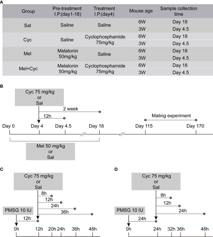
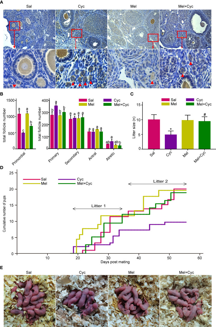
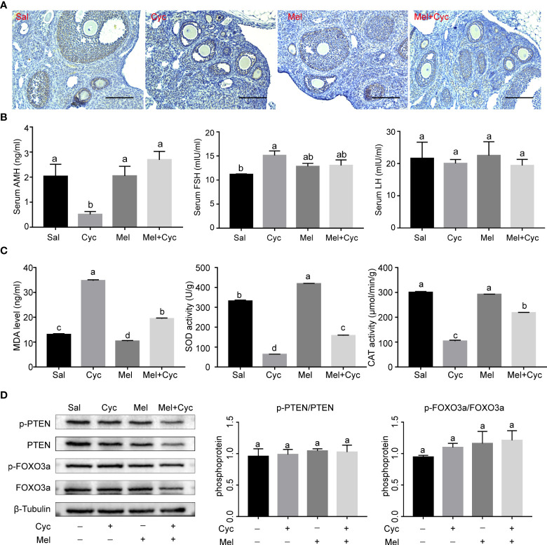
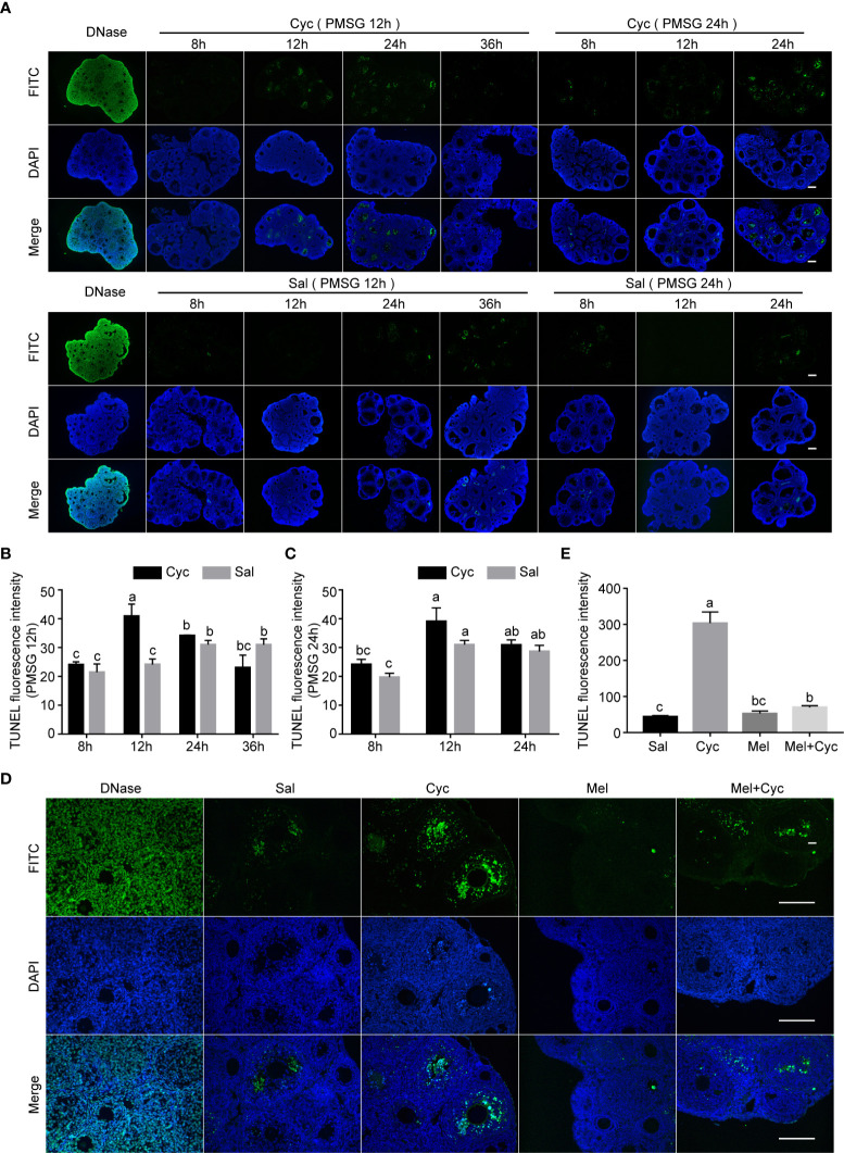
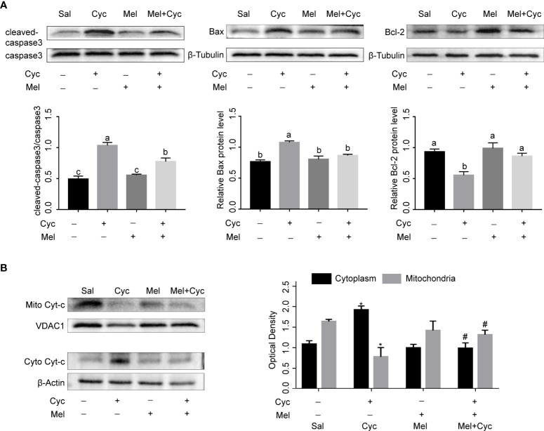
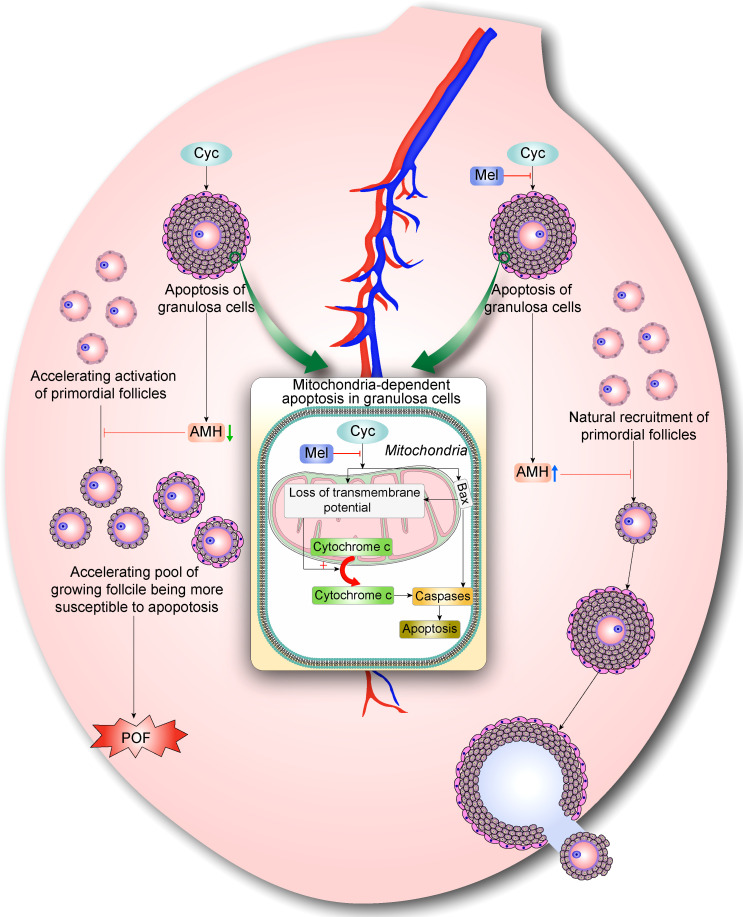
Cyclophosphaty -45mide (Cyc) chemotherapy in young female cancer patients is associated with an increased risk of premature ovarian insufficiency (POI). This study was designed to investigate the protective role of melatonin (Mel) as an adjuvant against Cyc-induced POI. Female mice received a single intraperitoneal (i.p.) dose of Cyc (75 mg/kg). Mel protection was achieved in mice after i.p. injection of melatonin (50 mg/kg) every 24 h for four consecutive days prior to chemotherapy initiation and for 14 additional days. Ovarian reserve testing, hormonal assays for follicle-stimulating hormone, luteinizing hormone, and anti-Müllerian hormone (AMH), assessment of the oxidative stress status, and measurement of the relative expression of genes in PTEN/AKT/FOXO3a and mitochondrial apoptosis pathways were performed. The results showed that treatment with 50 mg/kg Mel significantly prevented Cyc-induced over-activation of primordial follicles by maintaining the plasma level of AMH and subsequently preventing litter size reduction in mice treated with Cyc chemotherapy. Importantly, Mel treatment significantly prevented ovarian granulosa cell loss by inhibiting the mitochondrial apoptotic pathway. Identifying the protective actions of Mel against Cyc-induced primordial follicle loss has important implications for fertility maintenance in young cancer patients undergoing chemotherapy.

Keywords: anti-Mullerian hormone; apoptosis; cyclophosphamide; granulosa cell; melatonin; primordial follicle.


Conflict of interest statement

The authors declare that the research was conducted in the absence of any commercial or financial relationships that could be construed as a potential conflict of interest.

Figures

Figure 1
Figure 1
Graphic illustration of experimental schedule. (A) Mice were randomly divided into four groups. (B) Six or three-week-old ICR mice were pre-treated with i.p. injections of Mel or Sal once daily (at 24 h intervals) for four consecutive days before achieving Cyc injection, and for 14 additional days during chemotherapy treatment. On Day 4, mice were injected with Sal or Cyc (75 mg/kg). Blood and/or ovaries were collected 12 h or two weeks after Cyc injection. Fertility assessment was performed 115 days after Cyc injection. (C) Three-week-old prepubertal female mice were injected i.p. with 75 mg/kg body weight Cyc 12 h after injection of PMSG. Mice were sacrificed 8, 12, 24, or 36 h after chemotherapy treatment and their ovaries were excised (n = 4). (D) Prepubertal female mice were injected i.p. with 75 mg/kg body weight Cyc 24 h after injection of PMSG. Mice were sacrificed 12, 24, or 36 h after chemotherapy treatment and their ovaries were excised.
Figure 2
Figure 2
Mel prevents Cyc-induced primordial follicle loss. There were 25 six-week-old female mice in each group, of which 18 were sacrificed to obtain the ovary and blood samples, and seven mice were used for the mating experiment. (A) Oocytes in mouse ovaries were detected using an Nobox antibody (n = 6). Red arrow indicates primordial follicles. Bar = 200 μm. (B) Follicle count of whole ovarian tissue in control and treatment group mice 14 days after Cyc chemotherapy (n = 6). Mel prevented Cyc-induced dormant primordial follicle loss and suppressed the activation of primordial follicles into primary follicles. a,b,c,d Values with different letters in the same type of follicles were significantly different from each other. (C) Cyc treatment reduced the mean litter size in mice and Mel reversed this effect (n = 7). *P < 0.05 compared with Sal group. # P < 0.05 compared with Cyc group. (D) Cumulative pup number of each mouse in control and treatment groups at Day 55 of mating (n = 7). (E) Representative litters from control and treatment groups.
Figure 3
Figure 3
Effect of Mel on expression of AMH and oxidative stress in ovaries of six-week-old female mice 14 days after Cyc chemotherapy (Day 18). (A) AMH was expressed in granulosa cells of secondary and early antral follicles in Sal, Cyc, Mel, and Mel + Cyc groups (n = 6). Bar = 200 μm. (B) Cyc combined with Mel therapy prevented the decrease of serum AMH and LH levels but there was no significant change in serum FSH levels (n = 6). (C) Biochemical analysis of the ovarian CAT, SOD activities, and MDA levels (n = 6). (D) Phosphorylation levels of PTEN and FOXO3a did not change significantly after treatment with Cyc and Mel (n = 6). a,b Values with different letters are significantly different from each other.
Figure 4
Figure 4
Cyc induced apoptosis of granulosa cells in growing follicles and Mel prevented this apoptosis. (A) Three-week-old ICR mice were injected i.p. with Cyc 12 or 24 h after injection of PMSG, and apoptosis in ovarian tissues was detected by TUNEL kit at 8, 12, 24, and 36 h after Cyc injection. Regardless of whether Cyc was injected 12 or 24 h after PMSG injection, apoptotic peak of granulosa cells in ovary appeared 12 h after Cyc intervention, which was not related to the time of PMSG administration. Mice in control group were injected with Sal instead of Cyc, and the apoptotic peak of granulosa cells appeared 48 h (PMSG 12 h + Sal 36 h, or PMSG 24 h + Sal 24 h) after injection of PMSG. DNase I treatment was used for positive control of TUNEL assay. There were four three-week-old female mice in each group; Bar = 200 μm. (B) TUNEL fluorescence intensity in ovaries of 3-week-old ICR mice 8, 12, 24 and 36 hours after Cyc injection. The mice received Cyc chemotherapy 12 hours after injection of PMSG. (C) TUNEL fluorescence intensity in ovaries of 3-week-old ICR mice 8, 12 and 24 hours after Cyc injection. The mice received Cyc chemotherapy 24 hours after injection of PMSG. (D) Mel prevented apoptosis of granulosa cells 12 h after Cyc chemotherapy. There were four three-week-old female mice in each group; Bar = 200 μm. (E) TUNEL fluorescence intensity in ovaries of 3-week-old ICR mice in Sal, Cyc, Mel and Mel+Cyc groups 12 hours after Cyc injection.
Figure 5
Figure 5
Mel inhibits Cyc-induced apoptosis of granulosa cells through mitochondrial apoptosis pathway. (A) Western blot analysis was performed on cleaved-caspase3, caspase3, Bax, and Bcl-2 in the ovaries of three-week-old ICR mice 12 h after Cyc (75 mg/kg) treatment with or without Mel (n = 6). a,b,c Bars with different letters are significantly different in each group (P < 0.05). (B) Western blot analysis was performed on mitochondrial Cyt-c (mito Cyt-c) and cytoplasm Cyt-c (cyto Cyt-c) in the ovaries of three-week-old ICR mice 12 h after Cyc (75 mg/kg) treatment with or without Mel (n = 6). *P < 0.05 compared with Sal group. # P < 0.05 compared with Cyc group.
Figure 6
Figure 6
Mel prevents Cyc-induced over-activation of primordial follicles through inhibiting ovarian granulosa cell apoptosis and maintaining AMH expression. During normal follicle development, very few primordial follicles are selected and activated, whereas the vast majority of primordial follicles are maintained in a dormant state. AMH is mainly produced by granulosa cells in secondary follicles and early antral follicles. AMH can inhibit the activation of primordial follicles during recruitment and inhibit the sensitivity of antral follicles to FSH during recruitment cycle, thus maintaining AMH levels and selective activation of primordial follicle pool. Cyc exposure disturbs the balance of follicle activation by increasing apoptosis of AMH-secreting granulosa cells in growing follicles, resulting in further decrease of AMH levels and indirect over-activation of primordial follicle pool. Eventually, activated follicles undergo apoptosis and more primordial follicles are stimulated to become activated primary follicles, resulting in POF. With Mel co-treatment, activation of primordial follicles is reduced and apoptosis of AMH-secreting granulosa cells in growing follicles is decreased. Surviving granulosa cells maintained normal levels of AMH production, which regulates natural recruitment of primordial follicles and oogenesis. Hence, Mel restores the balance of follicle activation and returns ovaries to a healthy state.

Similar articles

Cited by

References

    1. Tarasiewicz M, Martynowicz I, Knapp P, Sieczynski P. "Oncofertility" procedures in children and adolescents. Pediatr Endocrinol Diabetes Metab (2019) 25(3):144–9. doi: 10.5114/pedm.2019.87710 - DOI - PubMed
    1. Goncalves V. Decisional regret in female oncofertility decision making-an integrative narrative review. Cancers (Basel) (2021) 13(19):1–15. doi: 10.3390/cancers13194735 - DOI - PMC - PubMed
    1. Devine PJ, Perreault SD, Luderer U. Roles of reactive oxygen species and antioxidants in ovarian toxicity. Biol Reprod (2012) 86(2):27. doi: 10.1095/biolreprod.111.095224 - DOI - PMC - PubMed
    1. Jang H, Hong K, Choi Y. Melatonin and fertoprotective adjuvants: Prevention against premature ovarian failure during chemotherapy. Int J Mol Sci (2017) 18(6):1–19. doi: 10.3390/ijms18061221 - DOI - PMC - PubMed
    1. Waxman J. Chemotherapy and the adult gonad: a review. J R Soc Med (1983) 76(2):144–8. doi: 10.1177/014107688307600212 - DOI - PMC - PubMed

Publication types