

Prenatal androgen treatment impairs the suprachiasmatic nucleus arginine-vasopressin to kisspeptin neuron circuit in female mice
- PMID: 35992143
- PMCID: PMC9388912
- DOI: 10.3389/fendo.2022.951344
Prenatal androgen treatment impairs the suprachiasmatic nucleus arginine-vasopressin to kisspeptin neuron circuit in female mice
Abstract
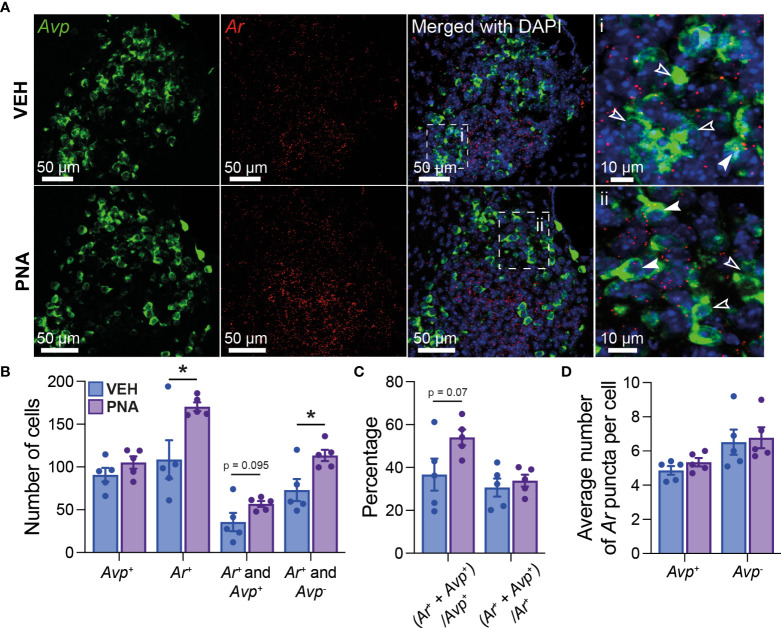
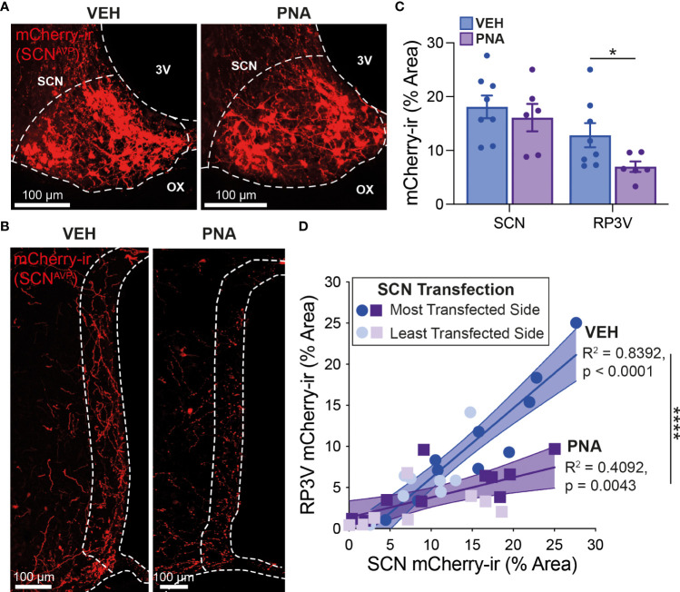
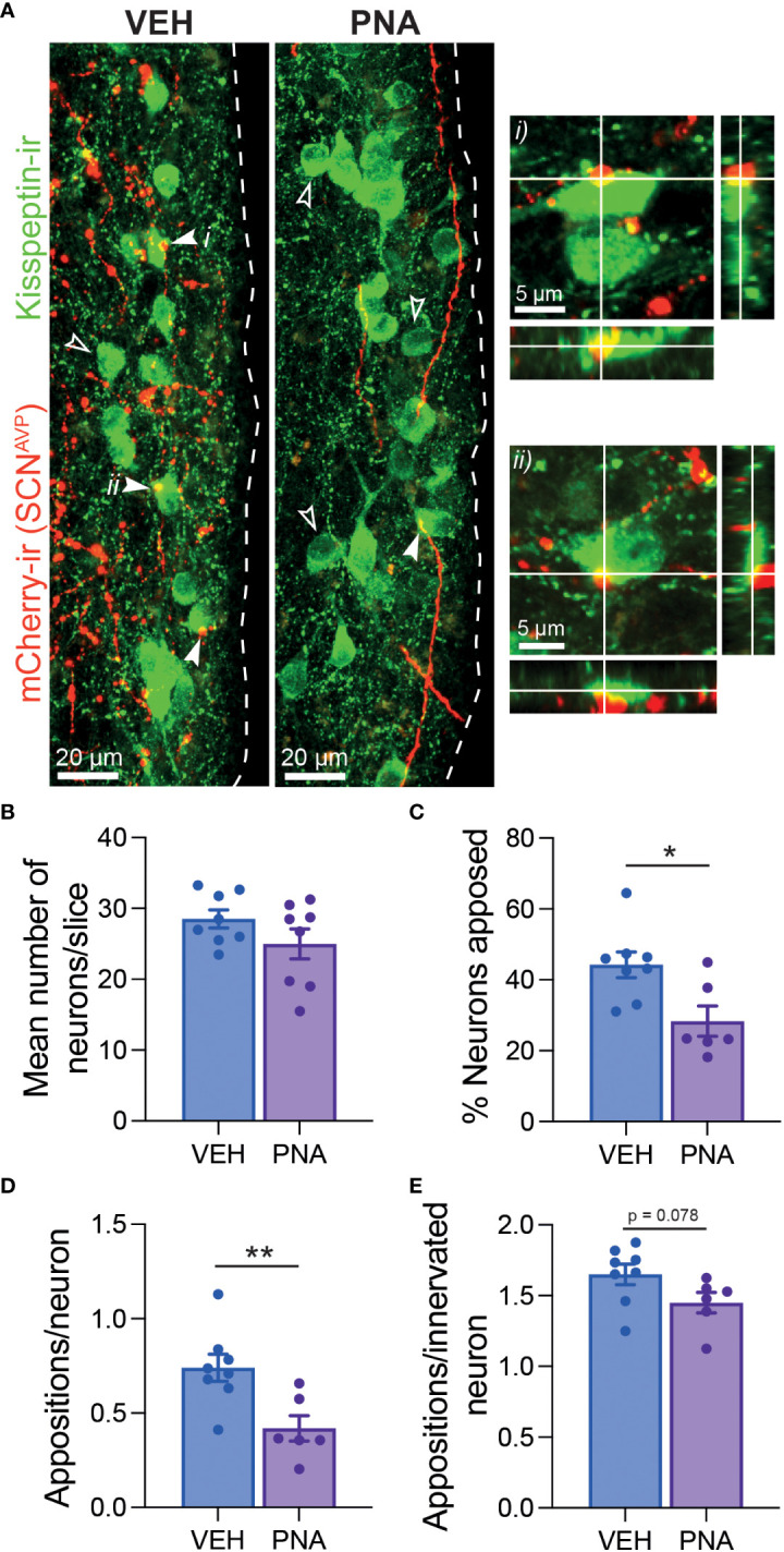
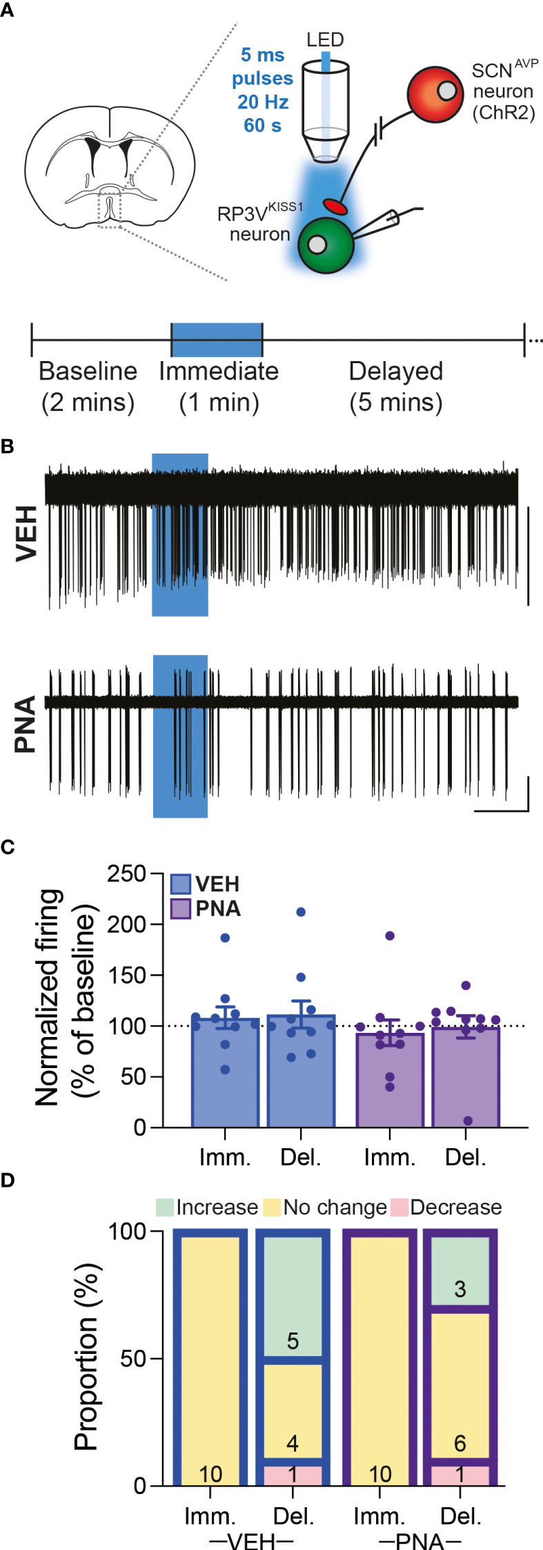
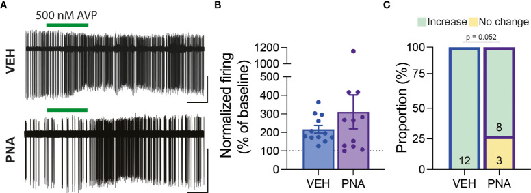
Polycystic ovary syndrome (PCOS) is associated with elevated androgen and luteinizing hormone (LH) secretion and with oligo/anovulation. Evidence indicates that elevated androgens impair sex steroid hormone feedback regulation of pulsatile LH secretion. Hyperandrogenemia in PCOS may also disrupt the preovulatory LH surge. The mechanisms through which this might occur, however, are not fully understood. Kisspeptin (KISS1) neurons of the rostral periventricular area of the third ventricle (RP3V) convey hormonal cues to gonadotropin-releasing hormone (GnRH) neurons. In rodents, the preovulatory surge is triggered by these hormonal cues and coincident timing signals from the central circadian clock in the suprachiasmatic nucleus (SCN). Timing signals are relayed to GnRH neurons, in part, via projections from SCN arginine-vasopressin (AVP) neurons to RP3VKISS1 neurons. Because rodent SCN cells express androgen receptors (AR), we hypothesized that these circuits are impaired by elevated androgens in a mouse model of PCOS. In prenatally androgen-treated (PNA) female mice, SCN Ar expression was significantly increased compared to that found in prenatally vehicle-treated mice. A similar trend was seen in the number of Avp-positive SCN cells expressing Ar. In the RP3V, the number of kisspeptin neurons was preserved. Anterograde tract-tracing, however, revealed reduced SCNAVP neuron projections to the RP3V and a significantly lower proportion of RP3VKISS1 neurons with close appositions from SCNAVP fibers. Functional assessments showed, on the other hand, that RP3VKISS1 neuron responses to AVP were maintained in PNA mice. These findings indicate that PNA changes some of the neural circuits that regulate the preovulatory surge. These impairments might contribute to ovulatory dysfunction in PNA mice modeling PCOS.
Keywords: GnRH; LH surge; PCOS; androgen receptor; circadian; electrophysiology; tract-tracing.
Copyright © 2022 Jamieson, Moore, Lohr, Thomas, Coolen, Lehman, Campbell and Piet.
Conflict of interest statement
The authors declare that the research was conducted in the absence of any commercial or financial relationships that could be construed as a potential conflict of interest.
Figures
Similar articles
-
Estrous Cycle Plasticity in the Central Clock Output to Kisspeptin Neurons: Implications for the Preovulatory Surge.Endocrinology. 2021 Jun 1;162(6):bqab071. doi: 10.1210/endocr/bqab071. Endocrinology. 2021. PMID: 33824970
-
Effects of electroacupuncture on the kisspeptin-gonadotropin-releasing hormone (GnRH) /luteinizing hormone (LH) neural circuit abnormalities and androgen receptor expression of kisspeptin/neurokinin B/dynorphin neurons in PCOS rats.J Ovarian Res. 2023 Jan 17;16(1):15. doi: 10.1186/s13048-022-01078-x. J Ovarian Res. 2023. PMID: 36650561 Free PMC article.
-
Circadian Function in Multiple Cell Types Is Necessary for Proper Timing of the Preovulatory LH Surge.J Biol Rhythms. 2019 Dec;34(6):622-633. doi: 10.1177/0748730419873511. Epub 2019 Sep 17. J Biol Rhythms. 2019. PMID: 31530063 Free PMC article.
-
The role of kisspeptin and RFamide-related peptide-3 neurones in the circadian-timed preovulatory luteinising hormone surge.J Neuroendocrinol. 2012 Jan;24(1):131-43. doi: 10.1111/j.1365-2826.2011.02162.x. J Neuroendocrinol. 2012. PMID: 21592236 Free PMC article. Review.
-
The role of kisspeptin in the pathogenesis of a polycystic ovary syndrome.Endocr Regul. 2023 Dec 21;57(1):292-303. doi: 10.2478/enr-2023-0032. Print 2023 Jan 1. Endocr Regul. 2023. PMID: 38127687 Review.
Cited by
-
Synergistic transcriptomic and metabolomic analyses in Zi geese ovaries with different clutch lengths.Poult Sci. 2025 Jul;104(7):105210. doi: 10.1016/j.psj.2025.105210. Epub 2025 Apr 23. Poult Sci. 2025. PMID: 40294555 Free PMC article.
-
Serotonin stimulates female preoptic area kisspeptin neurons via activation of type 2 serotonin receptors in mice.Front Endocrinol (Lausanne). 2023 Oct 12;14:1212854. doi: 10.3389/fendo.2023.1212854. eCollection 2023. Front Endocrinol (Lausanne). 2023. PMID: 37900129 Free PMC article.
-
Maternal androgen excess significantly impairs sexual behavior in male and female mouse offspring: Perspective for a biological origin of sexual dysfunction in PCOS.Front Endocrinol (Lausanne). 2023 Feb 15;14:1116482. doi: 10.3389/fendo.2023.1116482. eCollection 2023. Front Endocrinol (Lausanne). 2023. PMID: 36875467 Free PMC article.
-
Implication of vasopressin receptor genes (AVPR1A and AVPR1B) in the susceptibility to polycystic ovary syndrome.J Ovarian Res. 2024 Nov 5;17(1):214. doi: 10.1186/s13048-024-01515-z. J Ovarian Res. 2024. PMID: 39501331 Free PMC article.
References
MeSH terms
Substances
Grants and funding
LinkOut - more resources
Full Text Sources
Medical
Molecular Biology Databases
Research Materials
Miscellaneous