. 2022 Aug 3:13:902737.
doi: 10.3389/fendo.2022.902737. eCollection 2022.

Regulation of the kiss2 promoter in yellowtail clownfish (Amphiprion clarkii) by cortisol via GRE-dependent GR pathway

Affiliations

Regulation of the kiss2 promoter in yellowtail clownfish (Amphiprion clarkii) by cortisol via GRE-dependent GR pathway

Shao-Yang Bu et al. Front Endocrinol (Lausanne). .

Abstract

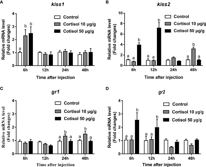
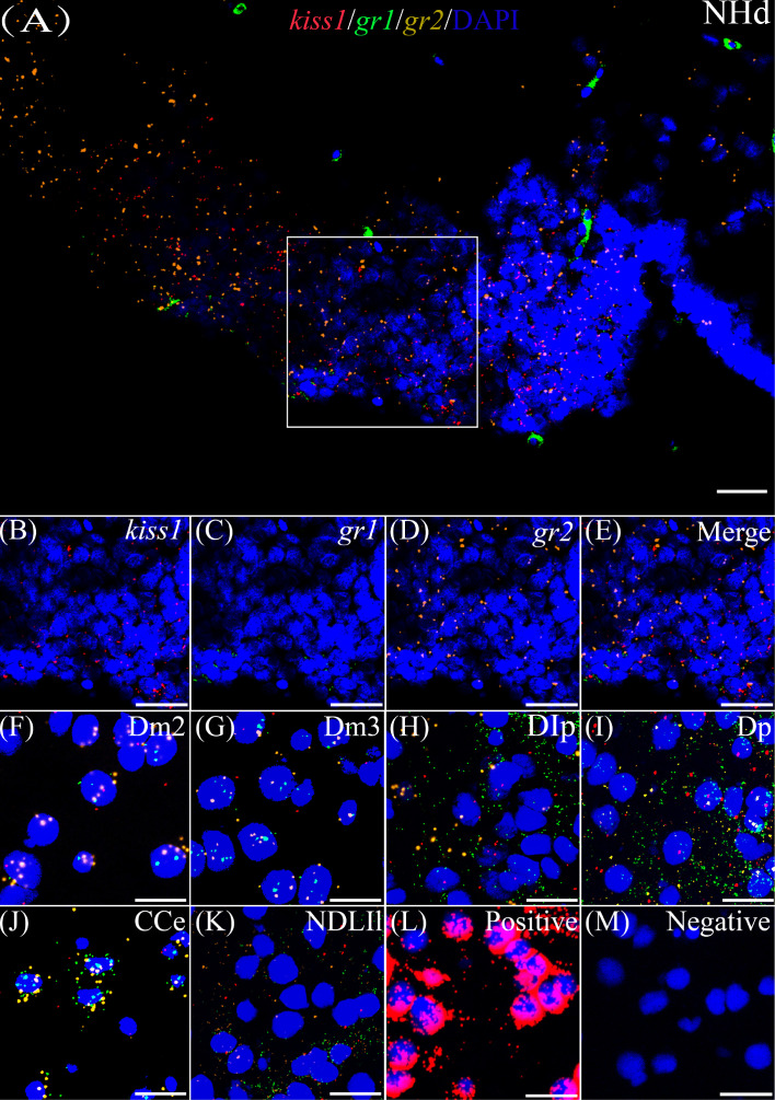
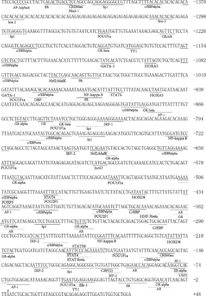
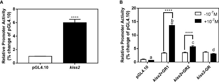
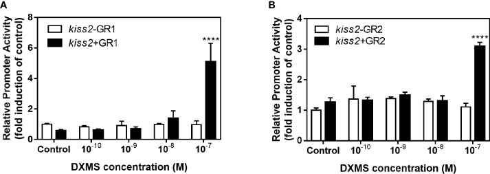
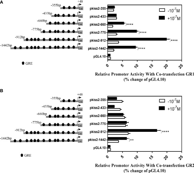
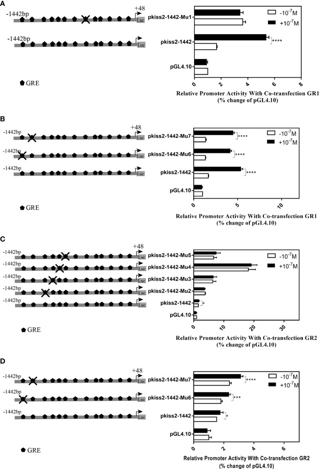
Kisspeptin plays a vital role in mediating the stress-induced reproductive regulation. Cortisol, known as a stress-related hormone, is involved in gonadal development and sexual differentiation by binding with glucocorticoid receptor (GR) to regulate the expression of kiss gene. In the present study, cortisol treatment in yellowtail clownfish (Amphiprion clarkii) showed that the expression of kiss (kiss1 and kiss2) and gr (gr1 and gr2) genes were increased significantly. We demonstrated that the yellowtail clownfish Kiss neurons co-express the glucocorticoid receptors in the telencephalon, mesencephalon, cerebellum, and hypothalamus. We further cloned the promoter of kiss2 gene in yellowtail clownfish and identified the presence of putative binding sites for glucocorticoid receptors, estrogen receptors, androgen receptors, progesterone receptors, AP1, and C/EBP. Applying transient transfection in HEK293T cells of the yellowtail clownfish kiss2 promoter, cortisol (dexamethasone) treatment was shown to enhance the promoter activities of the yellowtail clownfish kiss2 gene in the presence of GRs. Deletion analysis of kiss2 promoter indicated that cortisol-induced promoter activities were located between position -660 and -433 with GR1, and -912 and -775 with GR2, respectively. Finally, point mutation studies on the kiss2 promoter showed that cortisol-stimulated promoter activity was mediated by one GRE site located at position -573 in the presence of GR1 and by each GRE site located at position -883, -860, -851, and -843 in the presence of GR2. Results of the present study provide novel evidence that cortisol could regulate the transcription of kiss2 gene in the yellowtail clownfish via GRE-dependent GR pathway.

Keywords: Amphiprion clarkii; cortisol; glucocorticoid receptor; kiss2 promoter; stress.


Conflict of interest statement

The authors declare that the research was conducted in the absence of any commercial or financial relationships that could be construed as a potential conflict of interest.

Figures

Figure 1
Figure 1
Effects of cortisol on kiss1, kiss2, gr1, and gr2 mRNA levels in the brain of yellowtail clownfish. Expression of kiss1 (A), kiss2 (B), gr1 (C), and gr2 (D) after injection of cortisol with both concentrations at 10 and 50 μg/g during 48 h. The data are expressed as mean ± SEM (n = 5–8). Bars with different letters indicate significant differences between treatments at the same sampling time (p < 0.05).
Figure 2
Figure 2
Co-expression of gr1, gr2, and kiss1. The view of NHd (A) shows kiss1 (red), gr1 (green), and gr2 (yellow) with DAPI cell nuclear staining (blue). (B–E) The view of boxed region in panel (A), showing kiss1 (red), gr1 (green), and gr2 (yellow) with DAPI cell nuclear staining (blue) in panels (B–D), respectively, and the “merge” in panel (E) shows kiss1 (red), gr1 (green), and gr2 (yellow) with DAPI cell nuclear staining (blue). Representative images display gr1 (green) and gr2 (yellow) co-expression of kiss1 (red) with DAPI cell nuclear staining (blue) in Dm2 (F), Dm3 (G), DIp (H), Dp (I), CCe (J), and NDLIl (K). Positive and negative controls are shown in panels (L, M), respectively. NHd, dorsal habenular nucleus; Dm2, subdivision 2 of the medial dorsal telencephalic area; Dm3, subdivision 3 of the medial dorsal telencephalic area; DIp, lateral posterior part of the dorsal telencephalic area; Dp, posterior portion of the dorsal telencephalon; CCe, corpus of the cerebellum; NDLIl, lateral part of the diffuse nucleus of the inferior lobe. Bars = 20 μm.
Figure 3
Figure 3
Co-expression of gr1, gr2, and kiss2. The view of NRLd (A) shows kiss2 (red), gr1 (green), and gr2 (yellow) with DAPI cell nuclear staining (blue). (B–E) The view of boxed region in panel (A), showing kiss2 (red), gr1 (green), and gr2 (yellow) with DAPI cell nuclear staining (blue) in panels (B–D), respectively; and the “merge” in panel (E) shows kiss2 (red), gr1 (green), and gr2 (yellow) with DAPI cell nuclear staining (blue). Representative images display gr1 (green) and gr2 (yellow) co-expression of kiss2 (red) with DAPI cell nuclear staining (blue) in Dm2 (F), Dm3 (G), DIp (H), Dp (I), CCe (J), OT (K), NDLIl (L), NGp (M), TEG (N), and TPp (O). Positive and negative controls are shown in panels (P, Q), respectively. NRLd, dorsal part of the nucleus of the lateral recess; Dm2, subdivision 2 of the medial dorsal telencephalic area; Dm3, subdivision 3 of the medial dorsal telencephalic area; DIp, lateral posterior part of the dorsal telencephalic area; Dp, posterior portion of the dorsal telencephalon; CCe, corpus of the cerebellum; OT, optic tectum; NDLIl, lateral part of the diffuse nucleus of the inferior lobe; NGp, posterior part of glomerular nucleus; TEG, tegmentum; TPp, periventricular nucleus of the posterior tuberculum. Bars = 20 μm.
Figure 4
Figure 4
Sequence analysis of 5′-flanking region for kiss2. The numbering of the sequence is relative to the transcription start site marked on # and designated as +1. Putative binding sites for transcription factors are underlined and labeled. Transcription factor binding sites were predicted using the online PROMO and gene-regulation tool.
Figure 5
Figure 5
Effects of cortisol on kiss2 promoter activity. (A) Basic activity of kiss2 gene promoter in HEK-293T cell lines. The cells were transfected with 0.5 μg pkiss2-1442 and 0.025 μg pRL-CMV, or 0.5 μg pGL4.10 and 0.025 μg pRL-CMV as the control. Luciferase activity was measured after 24 h. Relative promoter activities are expressed as percentage of pGL4.10. (B) The activities of kiss2 promoter in the presence of cortisol in HEK293T cell lines. Cells were co-transfected with 0.5 μg pkiss2-1442, and 0.025 μg pRL-CMV with or without 0.05 μg yellowtail clownfish glucocorticoid receptor (GR1 or GR2) expression plasmid. The transfected cells were treated with or without 10−7 M cortisol. The luciferase activity was measured 24 h later. Relative promoter activities are expressed as percentage of pGL4.10 in the absence of cortisol. Data are represented as mean ± SEM (n = 4). **** (p < 0.0001) indicates that significant difference compared with the corresponding control. The different letters mean significant differences between groups with cortisol treatment (p < 0.05).
Figure 6
Figure 6
Effects of cortisol with different concentrations on the activities of kiss2 gene promoter. HEK-293T cells were transfected with 0.5 μg pkiss2-1442, 0.05 μg GR1 (A) or GR2 (B) and 0.025 μg pRL-CMV; 0.5 μg pGL4.10 co-transfected with 0.025 μg pRL-CMV as the control. Cells were treated with or without cortisol. Luciferase activity was detected after 24 h. Data are represented as mean ± SEM (n = 4). **** (p < 0.0001) indicates significant differences between groups with cortisol treatment.
Figure 7
Figure 7
Deletion analysis of the kiss2 promoter. Schematic of the putative GRE sites and deletion constructs of kiss2 promoter is shown on the left. HEK-293T cells were transfected with deletion constructs and pRL-CMV with GR1 (A) or GR2 (B) expression plasmid. Cells were incubated with or without 10−7 M cortisol treatment. Relative promoter activities are expressed as percentage of pGL4.10 in the absence of cortisol. Data are represented as mean ± SEM (n = 4). **(p < 0.01) and **** (p < 0.0001) indicate the significant differences compared with the corresponding control.
Figure 8
Figure 8
Mutations of the putative glucocorticoid receptor binding sites in kiss2 promoter. Schematic of mutated GRE sites is shown on the left. HEK-293T cells were transfected with GRE-mutated promoter constructs and pRL-CMV with GR1 (A, B) or GR2 (C, D) expression plasmid. Cells were incubated with or without 10−7 M cortisol treatment. Relative promoter activities are expressed as percentage of pGL4.10 in the absence of cortisol. Data are represented as mean ± SEM (n = 4). * (p < 0.05), *** (p < 0.001), and **** (p < 0.0001) indicate significant differences compared with the corresponding control.

Similar articles

References

    1. Castellano J, Roa J, Luque R, Dieguez C, Aguilar E, Pinilla L, et al. . KiSS-1/kisspeptins and the metabolic control of reproduction: physiologic roles and putative physiopathological implications. Peptides (2009) 30:139–45. doi: 10.1016/j.peptides.2008.06.007 - DOI - PubMed
    1. Ogawa S, Parhar IS. Anatomy of the kisspeptin systems in teleosts. Gen Comp Endocrinol (2013) 181:169–74. doi: 10.1016/j.ygcen.2012.08.023 - DOI - PubMed
    1. De Roux N, Genin E, Carel J-C, Matsuda F, Chaussain J-L, Milgrom E. Hypogonadotropic hypogonadism due to loss of function of the KiSS1-derived peptide receptor GPR54. Proc Natl Acad Sci (2003) 100:10972–6. doi: 10.1073/pnas.1834399100 - DOI - PMC - PubMed
    1. Seminara SB, Messager S, Chatzidaki EE, Thresher RR, Acierno JS, Jr, Shagoury JK, et al. . The GPR54 gene as a regulator of puberty. Obstetrical Gynecol Survey (2004) 59:351–3. doi: 10.1056/NEJMoa035322 - DOI - PubMed
    1. Hussain MA, Song W-J, Wolfe A. There is kisspeptin–and then there is kisspeptin. Trends Endocrinol Metab (2015) 26:564–72. doi: 10.1016/j.tem.2015.07.008 - DOI - PMC - PubMed

LinkOut - more resources