Rabbit hemorrhagic disease virus VP60 protein expressed in recombinant swinepox virus self-assembles into virus-like particles with strong immunogenicity in rabbits
- PMID: 35992711
- PMCID: PMC9387593
- DOI: 10.3389/fmicb.2022.960374
Rabbit hemorrhagic disease virus VP60 protein expressed in recombinant swinepox virus self-assembles into virus-like particles with strong immunogenicity in rabbits
Abstract
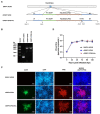
Rabbit Hemorrhagic Disease (RHD) is an economically significant infectious disease of rabbits, and its infection causes severe losses in the meat and fur industry. RHD Virus (RHDV) is difficult to proliferate in cell lines in vitro, which has greatly impeded the progress of investigating its replication mechanism and production of inactivated virus vaccines. RHDV VP60 protein is a major antigen for developing RHD subunit vaccines. Herein, we constructed a TK-deactivated recombinant Swinepox virus (rSWPV) expressing VP60 protein and VP60 protein coupled with His-tag respectively, and the expression of foreign proteins was confirmed using immunofluorescence assay and western blotting. Transmission electron microscopy showed that the recombinant VP60, with or without His-tag, self-assembled into virus-like particles (VLPs). Its efficacy was evaluated by comparison with available commercial vaccines in rabbits. ELISA and HI titer assays showed that high levels of neutralizing antibodies were induced at the first week after immunization with the recombinant strain and were maintained during the ongoing monitoring for the following 13 weeks. Challenge experiments showed that a single immunization with 106 PFU of the recombinant strain protected rabbits from lethal RHDV infection, and no histopathological changes or antigenic staining was found in the vaccine and rSWPV groups. These results suggest that rSWPV expressing RHDV VP60 could be an efficient candidate vaccine against RHDV in rabbits.
Keywords: VP60 protein; rabbit hemorrhagic disease virus; recombinant swinepox virus; vaccine; virus-like particles.
Copyright © 2022 Liu, Lin, Hu, Liu, Bian, Huang, Li, Yu, Luo and Deng.
Conflict of interest statement
FL was employed by Jiangxi Jinyibo Biotechnology Company. The remaining authors declare that the research was conducted in the absence of any commercial or financial relationships that could be construed as a potential conflict of interest.
Figures


Similar articles
-
The Baculovirus Expression System Expresses Chimeric RHDV VLPs as Bivalent Vaccine Candidates for Classic RHDV (GI.1) and RHDV2 (GI.2).Vaccines (Basel). 2025 Jun 27;13(7):695. doi: 10.3390/vaccines13070695. Vaccines (Basel). 2025. PMID: 40733672 Free PMC article.
-
Construction and immunogenicity of novel bivalent virus-like particles bearing VP60 genes of classic RHDV(GI.1) and RHDV2(GI.2).Vet Microbiol. 2020 Jan;240:108529. doi: 10.1016/j.vetmic.2019.108529. Epub 2019 Nov 30. Vet Microbiol. 2020. PMID: 31902498
-
DNA vaccination with a gene encoding VP60 elicited protective immunity against rabbit hemorrhagic disease virus.Vet Microbiol. 2013 May 31;164(1-2):1-8. doi: 10.1016/j.vetmic.2013.01.021. Epub 2013 Feb 1. Vet Microbiol. 2013. PMID: 23419819
-
Recombinant canine adenovirus type 2 expressing rabbit hemorrhagic disease virus VP60 protein provided protection against RHD in rabbits.Vet Microbiol. 2018 Jan;213:15-20. doi: 10.1016/j.vetmic.2017.11.007. Epub 2017 Nov 6. Vet Microbiol. 2018. PMID: 29291998
-
Adjuvant effects of interleukin-2 co-expression with VP60 in an oral vaccine delivered by attenuated Salmonella typhimurium against rabbit hemorrhagic disease.Vet Microbiol. 2019 Mar;230:49-55. doi: 10.1016/j.vetmic.2019.01.008. Epub 2019 Jan 8. Vet Microbiol. 2019. PMID: 30827404
Cited by
-
Swinepox virus: an unusual outbreak in free-range pig farms in Sicily (Italy).Porcine Health Manag. 2024 Jul 25;10(1):28. doi: 10.1186/s40813-024-00376-8. Porcine Health Manag. 2024. PMID: 39054554 Free PMC article.
-
Precocious Eimeria magna transgenically expressing RHDV P2 subdomain induces immune responses in rabbits.NPJ Vaccines. 2025 Jul 24;10(1):167. doi: 10.1038/s41541-025-01223-9. NPJ Vaccines. 2025. PMID: 40707472 Free PMC article.
-
First report of GI.1aP-GI.2 recombinants of rabbit hemorrhagic disease virus in domestic rabbits in China.Front Microbiol. 2023 Jul 14;14:1188380. doi: 10.3389/fmicb.2023.1188380. eCollection 2023. Front Microbiol. 2023. PMID: 37520350 Free PMC article.
-
The Baculovirus Expression System Expresses Chimeric RHDV VLPs as Bivalent Vaccine Candidates for Classic RHDV (GI.1) and RHDV2 (GI.2).Vaccines (Basel). 2025 Jun 27;13(7):695. doi: 10.3390/vaccines13070695. Vaccines (Basel). 2025. PMID: 40733672 Free PMC article.
References
-
- Barcena J., Morales M., Vazquez B., Boga J. A., Parra F., Lucientes J., et al. . (2000). Horizontal transmissible protection against myxomatosis and rabbit hemorrhagic disease by using a recombinant myxoma virus. J. Virol. 74, 1114–1123. doi: 10.1128/JVI.74.3.1114-1123.2000, PMID: - DOI - PMC - PubMed
LinkOut - more resources
Full Text Sources

