

Epithelial-myoepithelial carcinoma of the nasopharynx: A case report and review of the literature
- PMID: 35992786
- PMCID: PMC9389165
- DOI: 10.3389/fonc.2022.923579
Epithelial-myoepithelial carcinoma of the nasopharynx: A case report and review of the literature
Abstract
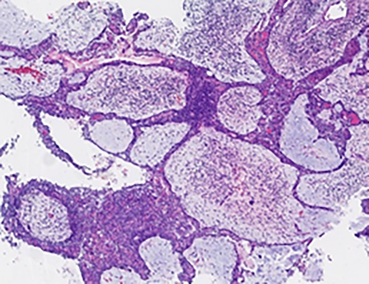
Background: Epithelial-myoepithelial carcinoma (EMCa) is a rare low-grade malignant tumor that most commonly occurs in the salivary glands, with approximately 320 cases having been reported worldwide. Here, we report the third case of EMCa occurring in the nasopharynx. Rare cases in the breast, pituitary gland, lacrimal gland, nose, paranasal sinus, nasal cavity, trachea and bronchus, lung, and even the pleura mediastinalis have also been reported. Histopathology and immunohistochemistry are useful for confirming the diagnosis of EMCa, which is characterized by biphasic tubular structures composed of inner ductal and outer clear myoepithelial cells and stains for different markers in each layer. However, because of the rarity of EMCa, the clinicopathological characteristics and treatment of these patients remain unclear.
Case presentation: We report a rare case of EMCa of the nasopharynx. A 51-year-old man presented with a 5-month history of pain while swallowing and aggravation accompanied by right ear tinnitus lasting for 1 month. Nasopharyngoscopy and magnetic resonance imaging (MRI) of the nasopharynx and neck revealed a 5.6 cm × 3.4 cm × 3.1 cm mass in the nasopharyngeal space, invasion of the right cavernous sinus, and lymph node enlargement in the right retropharyngeal space. On 17 April 2019, based on the histopathological and immunohistochemical features, a final diagnosis of EMCa of the right nasopharynx was made. The patient underwent concurrent chemoradiotherapy (CCRT), and his symptoms were relieved after treatment. On 10 January 2022, nasopharynx MRI and biopsy revealed local recurrence, but chest and abdominal computed tomography (CT) showed no obvious signs of metastasis. The local recurrence-free survival (LRFS) period was 33 months.
Conclusion: To the best of our knowledge, this is the third reported case of EMCa in the nasopharynx and the only case of EMCa in the nasopharynx treated with CCRT, and a partial response was achieved. Therefore, to improve the quality of life and prognosis of patients with unresectable tumors, we believe that CCRT is a suitable option. Further clinical observations are required to elucidate the pathophysiology and prognosis of EMCa.
Keywords: case report; concurrent chemoradiotherapy; epithelial–myoepithelial carcinoma; immunohistochemistry; nasopharynx.
Copyright © 2022 Zhang, Wang, Wang, Zhang, Li, Li, Cai, Ren, Zhang and Hao.
Conflict of interest statement
The authors declare that the research was conducted in the absence of any commercial or financial relationships that could be construed as a potential conflict of interest.
Figures


Similar articles
-
Epithelial-myoepithelial carcinoma arising in the nasal cavity: a case report and review of literature.Pathology. 1999 May;31(2):148-51. doi: 10.1080/003130299105340. Pathology. 1999. PMID: 10399171 Review.
-
Epithelial-myoepithelial carcinoma of the trachea-a rare entity case report.J Thorac Dis. 2014 Mar;6 Suppl 1(Suppl 1):S194-9. doi: 10.3978/j.issn.2072-1439.2013.11.17. J Thorac Dis. 2014. PMID: 24672694 Free PMC article.
-
Epithelial-myoepithelial carcinoma: a review of the clinicopathologic spectrum and immunophenotypic characteristics in 61 tumors of the salivary glands and upper aerodigestive tract.Am J Surg Pathol. 2007 Jan;31(1):44-57. doi: 10.1097/01.pas.0000213314.74423.d8. Am J Surg Pathol. 2007. PMID: 17197918
-
Epithelial-Myoepithelial Carcinoma of the Lacrimal Sac and Literature Review of the Lacrimal System.Allergy Rhinol (Providence). 2020 Apr 20;11:2152656720920600. doi: 10.1177/2152656720920600. eCollection 2020 Jan-Dec. Allergy Rhinol (Providence). 2020. PMID: 32341837 Free PMC article.
-
Oncocytic and apocrine epithelial myoepithelial carcinoma: novel variants of a challenging tumor.Head Neck Pathol. 2013 Jul;7 Suppl 1(Suppl 1):S77-84. doi: 10.1007/s12105-013-0461-0. Epub 2013 Jul 3. Head Neck Pathol. 2013. PMID: 23821213 Free PMC article. Review.
Cited by
-
Unusual pulmonary nodules diffusion from epithelial‑myoepithelial carcinoma on 18F‑FDG PET/CT: A case report.Oncol Lett. 2024 Oct 22;29(1):19. doi: 10.3892/ol.2024.14765. eCollection 2025 Jan. Oncol Lett. 2024. PMID: 39492931 Free PMC article.
-
Epstein-Barr virus-associated poorly differentiated nasopharyngeal adenocarcinoma: a case report and literature review.J Int Med Res. 2023 May;51(5):3000605231173839. doi: 10.1177/03000605231173839. J Int Med Res. 2023. PMID: 37203382 Free PMC article. Review.
References
-
- Seethala R, Barnes E, Hunt J. Epithelial-myoepithelial carcinoma: a review of the clinicopathologic spectrum and immunophenotypic characteristics in 61 tumors of the salivary glands and upper aerodigestive tract. Am J Surg Pathol (2007) 31(1):44–57. doi: 10.1097/01.pas.0000213314.74423.d8 - DOI - PubMed
Publication types
LinkOut - more resources
Full Text Sources
Miscellaneous