. 2022 Aug 3:12:939965.
doi: 10.3389/fonc.2022.939965. eCollection 2022.

Metastases to the thyroid gland: ultrasonographic findings and diagnostic value of fine-needle aspiration cytology

Affiliations

Metastases to the thyroid gland: ultrasonographic findings and diagnostic value of fine-needle aspiration cytology

Zhenyun Tang et al. Front Oncol. .

Abstract

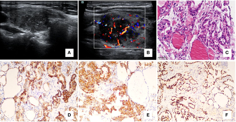
Introduction: In the present study, we aimed to analyze ultrasonographic findings of metastases to the thyroid and explore the role of fine-needle aspiration cytology (FNAC) in the diagnosis of metastases to the thyroid.

Methods: Twelve cases of cytologically or/and pathologically confirmed metastatic tumors of the thyroid gland were reviewed. All the primary thyroid lesions and lymphomas were excluded. The location, maximum size, echogenicity, shape, margin, presence of calcifications, vascularity, and cervical lymph nodes were assessed on ultrasonography. In addition, the results of cytology or pathology (or both) were noted retrospectively.

Results: Eight of 10 patients were diagnosed correctly with FNAC. Two cases presented with diffuse involvement in both thyroid lobes. Nine cases demonstrated a hypoechoic nodule with an irregular margin, four of which had microcalcifications. One case presented with a mixed solid and cystic mass with an oval shape. The lesions with cervical lymph nodes were found in seven cases.

Conclusion: Most metastatic thyroid cancer has similar ultrasound features to primary thyroid cancer. In some cases with atypical US features, ultrasound diagnosis should be combined with the medical history. FNAC might be helpful in the diagnosis.

Keywords: fine-needle aspiration cytology; metastasis to the thyroid gland; secondary thyroid neoplasm; thyroid; thyroid metastasis; ultrasound.


Conflict of interest statement

The authors declare that the research was conducted in the absence of any commercial or financial relationships that could be construed as a potential conflict of interest.

Figures

Figure 1
Figure 1
Case 5, a 69-year-old woman had a 7-year history of clear cell renal cell carcinoma (RCC). A regular and circumscribed nodule with solid and cystic composition was found on ultrasonography in the left thyroid lobe. The nodule showed a parallel orientation and no calcifications (A, B) with rich intra-tumoral vascularity (C). No suspicious cervical lymph nodes were detected on ultrasonography. The postoperative pathology showed metastatic RCC. Histology showed a well-demarcated expanding tumor with a fibrous capsule (D – 100x), which had the characteristics of RCC with delicate arborizing vessels and background colloid and typical vessel formation (E – 200x), large vacuolated clear cytoplasm, eosinophilic cytoplasm and mild nuclear atypia (F– 400x), (A–C) ultrasonography; (D–F) hematoxylin-eosin staining.
Figure 2
Figure 2
Case 4, a 51-year-old woman had a 3-year history of lung adenocarcinoma. Ultrasonography of the right lobe (A) showed an irregular, hypoechoic nodule with microcalcifications. Doppler ultrasonography showed rich intra-tumoral vascularity (B). The cytology result was atypia with an undetermined source, and then a core needle biopsy was performed. The histology showed the papillary and tubular-acinar architecture and malignant cuboidal to columnar tumor cells with mild nuclear atypia [(C) – 400x]. In immunohistochemistry, the malignant cells were positive for CK7 (D – 200x), Napsin A [(E) – 200x], and TTF1 (F – 200x). (A, B) ultrasonography; (C) hematoxylin-eosin staining; (D–F) immunohistochemistry.
Figure 3
Figure 3
Case12, a 68-year-old female patient had a 7-year history of malignant breast tumor. Ultrasonography showed an irregular and hypoechoic nodule with micro-calcifications (A). On color Doppler, it showed intra-nodular high vascularization (B). The patient underwent fine-needle aspiration cytology. The cell mass exhibited scattered malignant tumor cells with necrosis, large hyperchromatic nuclei, and irregular nuclear membranes [(C) – 200x]. In immunohistochemistry, the malignant cells showed negative expressions of estrogen receptor [(D) – 200x] and progesterone receptor [(E) – 200x], and positive for HER2 (F – 200x), GATA-3 [(G) – 200x], GALACTIN-3 (H – 200x), and AE1/AE3 ((I)– 200x). (A, B) ultrasonography; (C) hematoxylin-eosin staining; (D–I) immunohistochemistry.

References

    1. Papi G, Fadda G, Corsello SM, Corrado S, Rossi ED, Radighieri E, et al. . Metastases to the thyroid gland: prevalence, clinicopathological aspects and prognosis: a 10-year experience. Clin Endocrinol (Oxf) (2007) 66(4):565–71. doi: 10.1111/j.1365-2265.2007.02773.x - DOI - PubMed
    1. HooKim K, Gaitor J, Lin O, Reid MD. Secondary tumors involving the thyroid gland: A multi-institutional analysis of 28 cases diagnosed on fine-needle aspiration. Diagn Cytopathol (2015) 43(11):904–11. doi: 10.1002/dc.23331 - DOI - PubMed
    1. Russell JO, Yan K, Burkey B, Scharpf J. Nonthyroid metastasis to the thyroid gland: Case series and review with observations by primary pathology. Otolaryngol Head Neck Surg (2016) 155(6):961–8. doi: 10.1177/0194599816655783 - DOI - PubMed
    1. Cordes M, Kuwert T. Metastases of non-thyroidal tumors to the thyroid gland: a regional survey in middle franconia. Exp Clin Endocrinol Diabetes (2014) 122(5):273–6. doi: 10.1055/s-0034-1372623 - DOI - PubMed
    1. Cichoń S, Anielski R, Konturek A, Barczyński M, Cichoń W. Metastases to the thyroid gland: seventeen cases operated on in a single clinical center. Langenbecks Arch Surg (2006) 391(6):581–7. doi: 10.1007/s00423-006-0081-1 - DOI - PubMed

LinkOut - more resources