. 2022 Aug 5:12:933446.
doi: 10.3389/fonc.2022.933446. eCollection 2022.

Small molecule MMRi62 targets MDM4 for degradation and induces leukemic cell apoptosis regardless of p53 status

Affiliations

Small molecule MMRi62 targets MDM4 for degradation and induces leukemic cell apoptosis regardless of p53 status

Rati Lama et al. Front Oncol. .

Abstract

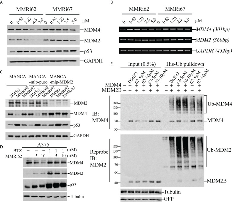
MDM2 and MDM4 proteins are key negative regulators of tumor suppressor p53. MDM2 and MDM4 interact via their RING domains and form a heterodimer polyubiquitin E3 ligase essential for p53 degradation. MDM4 also forms heterodimer E3 ligases with MDM2 isoforms that lack p53-binding domains, which regulate p53 and MDM4 stability. We are working to identify small-molecule inhibitors targeting the RING domain of MDM2-MDM4 (MMRi) that can inactivate the total oncogenic activity of MDM2-MDM4 heterodimers. Here, we describe the identification and characterization of MMRi62 as an MDM4-degrader and apoptosis inducer in leukemia cells. Biochemically, in our experiments, MMRi62 bound to preformed RING domain heterodimers altered the substrate preference toward MDM4 ubiquitination and promoted MDM2-dependent MDM4 degradation in cells. This MDM4-degrader activity of MMRi62 was found to be associated with potent apoptosis induction in leukemia cells. Interestingly, MMRi62 effectively induced apoptosis in p53 mutant, multidrug-resistant leukemia cells and patient samples in addition to p53 wild-type cells. In contrast, MMRi67 as a RING heterodimer disruptor and an enzymatic inhibitor of the MDM2-MDM4 E3 complex lacked MDM4-degrader activity and failed to induce apoptosis in these cells. In summary, this study identifies MMRi62 as a novel MDM2-MDM4-targeting agent and suggests that small molecules capable of promoting MDM4 degradation may be a viable new approach to killing leukemia cells bearing non-functional p53 by apoptosis.

Keywords: E3 ligase; MDM2; MDM4; apoptosis; degradation; leukemia; p53; ubiquitination.


Conflict of interest statement

The authors declare that the research was conducted in the absence of any commercial or financial relationships that could be construed as a potential conflict of interest.

Figures

Figure 1
Figure 1
Identification of MMRi62 as an apoptosis inducer and MMRi67 as E3 ligase inhibitor by apoptosis and E3 ligase inhibitor screens. (A) Apoptosis screen of MMRi6 analogs by activated caspase 3 (AC3) and cleaved PARP as readouts using wt-p53 bearing MV4-11 cells treated with 2 μM of MMRi6 analogs for 24 h. (B) In vitro ubiquitination assay using recombinant MDM2B, MDM4, and p53 proteins in the presence of the solvent dimethyl sulfoxide (DMSO) or MMRi6 analogs (0, 5, and 10 μM of MMRi62 or MMRi67); ubiquitinated species of MDM2B, MDM4, p53, and polyubiquitin are shown. (C) In vitro ubiquitination assays as performed in panel B using MDM4 and MDM2B with an extended range of concentrations of MMRi62 and MMRi67; ubiquitinated species of MDM2B and MDM4 are shown.
Figure 2
Figure 2
Characterization of drug–target interactions in vitro. (A) Left, in vitro pulldown assay with recombinant FLAG-MDM4 and HA-MDM2B in the presence of solvent dimethyl sulfoxide (DMSO) or 5 and 10 μM of MMRi62 or MMRi67. After anti-FLAG pulldown, the FLAG-MDM4-bound HA-MDM2B was detected by Western blotting (WB) with anti-HA antibody. Right, chemical structures of MMRi62 and MMRi67. (B) Measurement of the binding affinity of MMRi62 and MMRi67 to preformed RING–RING heterodimers of MDM2 and MDM4 in microscale thermophoresis (MST) analyses. MST analyses were performed in the presence of MMRi62 or MMRi67 at concentrations ranging from 3 nM to 100 μM obtained through serial dilutions. The fluorescence intensities (y-axis, Fnorm%) were normalized to the overall highest detected signal. The equilibrium dissociation constant between the MMRi62/MMRi67 and RING heterodimers (Kd) is presented. Top, the calculated Kd for MMRi62 and MMRi67; bottom, the fitting curve of the measurements using MO.Affinity Analysis software. (C) Molecular modeling of interaction interfaces for the RING domains of MDM2 and MDM4 with MMRi62 and MMRi67. Top, amino acid alignment of RING domains of MDM2 and MDM4. Middle, the interaction residues of RING domains with MMRi62 and MMRi67 identified by docking analysis. The F490 region in the MDM2 RING is critical for E3 ligase activity of MDM2, and the H456 region in the MDM4 RING is critical for chelating a Zn to maintain the MDM4 RING domain structure. Bottom, presentations of the docking interface—the results for the MMRi67R enantiomer are shown.
Figure 3
Figure 3
MMRi62 induces MDM2-dependent degradation of MDM4 protein in cells. (A) Western blotting analysis of the effects of MMRi62 and MMRi67 on expression levels of p53, MDM2, and MDM4 in NALM6 cells treated for 24 h at the indicated concentrations. (B) RT-PCR analysis of the effects of MMRi62 and MMRi67 on p53, Mdm2, and Mdm4 mRNA expression in the same NALM6 samples prepared in panel (A). (C) Results demonstrating that MDM2 is required for MMRi62-induced degradation of MDM4 proteins in cells. The Western blotting (WB) analysis was performed as in panel A except in MANCA cells, or cells stably expressing empty vector mlp (MANCA-mlp-puro) or stably expressing MDM2-mcRNA (MANCA-mlp-MDM2); cells were treated with 5 μM of MMRi62 or MMRi67. (D) Western blotting analysis of MDM4 degradation by MMRi62 rescued by proteasome inhibition with bortezomib (BTZ); in these experiments, A375 cells were treated with MMRi62 for 24 h with or without BTZ for 16 h before cell harvest. (E) MMRi62 induced increased ubiquitination of MDM4 and MDM2 in 293 cells during in vivo ubiquitination assays. The 293 cells were transfected with vectors expressing MDM2B, FLAG-MDM4, His-ubiquitin, and green fluorescent protein (GFP). Cell lysates were prepared 24 h after transfection and used for His-tag pulldown followed by WB with anti-FLAG (for MDM4) and anti-MDM2 (mAb 4B11). Ubiquitinated MDM4 and MDM2B are indicated. GFP was used for the internal control of transfection efficiency and protein inputs.
Figure 4
Figure 4
MMRi62 but not MMRi67 inhibited the proliferation of leukemic cells by inducing p53-independent apoptosis with low toxicity for healthy peripheral blood monocytes (PBMCs). (A) Growth curves of wt-p53 NALM6 cells in the presence of different concentrations of MMRi62 or MMRi67 in 72-h proliferation assays. (B) Growth curve of PBMCs in the presence of different concentrations of MMRi62 in comparison with that of NALM6 cells replotted from the same dataset in panel (A) PBMC proliferation was stimulated by 10 μg/ml of pokeweed mitogen (PWM). (C) Flow cytometry analysis of annexin-V-positive apoptotic cells after 48-h treatment with 5 mM of MMRi62 or MMRi67. (D) Concentration-dependent caspase-3 activation (AC3) and PARP cleavage (cPARP) in NALM6 cells and NALM6shp53 cells stably expressing shRNA against p53. The cells were treated with the indicated concentrations of either MMRi62 or MMRi67 for 24 h followed by Western blotting (WB) analysis with corresponding antibodies. (E) Similar analysis as in panel (D) except using p53-null HL60 cells.
Figure 5
Figure 5
Results demonstrating that MMRi62 is a potent apoptosis inducer in acquired drug-resistant leukemia cells. (A) Growth curves of p53-null HL60 and HL60VR cells in the presence of different concentrations of vincristine (left), daunorubicin (middle), and MMRi62 (right). IC50s of each drug for the two cell lines are shown beside the corresponding curves. (B) Flow cytometry analysis of annexin-V-positive apoptotic cells in non-treated cells (left) or cells treated with 5 uM of daunorubicin (middle) or 5µM of MMRi62 (right) for 48 h. (C) Western blotting analysis of MDM2 and MDM4 expression, caspase-3 activation, and apoptotic PARP cleavage in HL60VR cells treated for 24 h at the indicated concentrations of MMRi62 or daunorubicin (Daun). (D) Flow cytometry analysis of annexin-V-positive apoptotic cells after 72-h treatment using an acute myeloid leukemia (AML) patient sample (Patient#02-1919) with the indicated concentrations of MMRi62. (E) Quantitative graph of the apoptotic fractions in different treatments in panel (D).
Figure 6
Figure 6
MMRi62 induced p53-independent apoptosis and inhibited colony-forming unit (CFU) formation in primary acute myeloid leukemia (AML) patient samples in vitro. (A) Representative images of CFU plates taken after 13 days of culture with primary AML patient samples pre-treated with the indicated concentrations of MMRi62 or MMRi67 for 4 h in a 37°C–5% CO2 incubator followed by culturing them in fully supplemented CFU growth media. (B) Quantitative graph of CFU numbers for two AML patient samples treated with either MMRi62 or MMRi67 at different concentrations.

References

    1. Freireich EJ, Wiernik PH, Steensma DP. The leukemias: A half-century of discovery. J Clin Oncol (2014) 32(31):3463–9. doi: 10.1200/JCO.2014.57.1034 - DOI - PubMed
    1. Kantarjian HM, Keating MJ, Freireich EJ. Toward the potential cure of leukemias in the next decade. Cancer (2018) 124(22):4301–13. doi: 10.1002/cncr.31669 - DOI - PubMed
    1. Desikan SP, Daver N, DiNardo C, Kadia T, Konopleva M, Ravandi F. Resistance to targeted therapies: Delving into FLT3 and IDH. Blood Cancer J (2022) 12(6):91. doi: 10.1038/s41408-022-00687-5 - DOI - PMC - PubMed
    1. Arai Y, Chi S, Minami Y, Yanada M. FLT3-targeted treatment for acute myeloid leukemia. Int J Hematol (2022). doi: 10.1007/s12185-022-03374-0 - DOI - PubMed
    1. Montesinos P, Recher C, Vives S, Zarzycka E, Wang J, Bertani G, et al. . Ivosidenib and azacitidine in idh1-mutated acute myeloid leukemia. N Engl J Med (2022) 386(16):1519–31. doi: 10.1056/NEJMoa2117344 - DOI - PubMed