. 2022 Aug 5:16:972658.
doi: 10.3389/fnins.2022.972658. eCollection 2022.

Anterior cingulate cortex and its projections to the ventral tegmental area regulate opioid withdrawal, the formation of opioid context associations and context-induced drug seeking

Affiliations

Anterior cingulate cortex and its projections to the ventral tegmental area regulate opioid withdrawal, the formation of opioid context associations and context-induced drug seeking

Greer McKendrick et al. Front Neurosci. .

Abstract

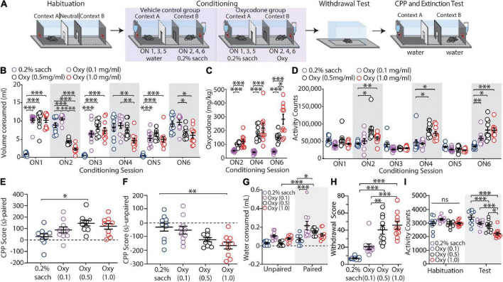
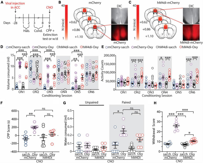
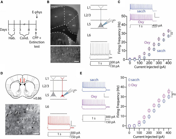
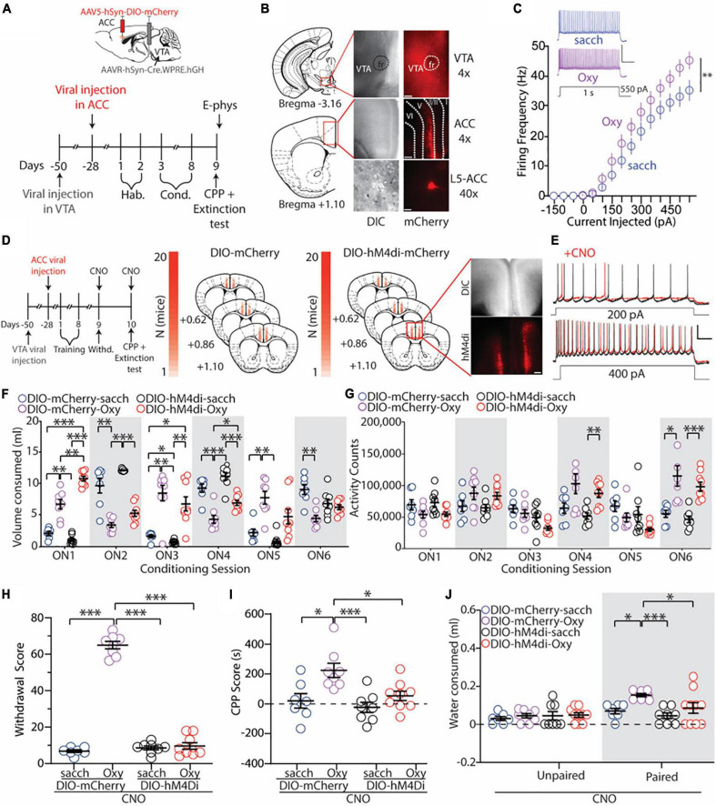
Clinical evidence suggests that there are correlations between activity within the anterior cingulate cortex (ACC) following re-exposure to drug-associated contexts and drug craving. However, there are limited data contributing to our understanding of ACC function at the cellular level during re-exposure to drug-context associations as well as whether the ACC is directly related to context-induced drug seeking. Here, we addressed this issue by employing our novel behavioral procedure capable of measuring the formation of drug-context associations as well as context-induced drug-seeking behavior in male mice (8-12 weeks of age) that orally self-administered oxycodone. We found that mice escalated oxycodone intake during the long-access training sessions and that conditioning with oxycodone was sufficient to evoke conditioned place preference (CPP) and drug-seeking behaviors. Additionally, we found that thick-tufted, but not thin-tufted pyramidal neurons (PyNs) in the ACC as well as ventral tegmental area (VTA)-projecting ACC neurons had increased intrinsic membrane excitability in mice that self-administered oxycodone compared to controls. Moreover, we found that global inhibition of the ACC or inhibition of VTA-projecting ACC neurons was sufficient to significantly reduce oxycodone-induced CPP, drug seeking, and spontaneous opioid withdrawal. These results demonstrate a direct role of ACC activity in mediating context-induced opioid seeking among other behaviors, including withdrawal, that are associated with the DSM-V criteria of opioid use disorder.

Keywords: CPP; anterior cingulate cortex; oxycodone; self-administration; ventral tegmental area.


Conflict of interest statement

The authors declare that the research was conducted in the absence of any commercial or financial relationships that could be construed as a potential conflict of interest.

Figures

FIGURE 1
FIGURE 1
Oxycodone oral self-administration evokes conditioned place preference, context-induced drug-seeking, and withdrawal. (A) Timeline of the behavioral procedure of saccharin control or oxycodone oral-SA-CPP. Mice underwent habituation sessions followed by 6 overnight conditioning sessions where they were given access to water (control) or saccharin (experimental) in the unpaired chambers and saccharin (control) or oxycodone diluted in saccharin solution (experimental) in the paired chambers. 1-2 days later, mice were given withdrawal testing followed by a 2 h extinction test. (B) Summary graph showing the volume of solution consumed between groups during overnight (ON) conditioning sessions 1-6 (ON 1-6) [F(15,180) = 29.84, p < 0.0001, two-way repeated-measures ANOVA, Bonferroni post hoc test]. (C) Summary graph showing the mg/kg of oxycodone consumed during ON 2, 4, and 6 [F(4,54) = 13.49, p < 0.001, two-way repeated-measures ANOVA, Bonferroni post hoc test]. (D) Summary graph showing the locomotor activity counts across conditioning sessions [F(15,180) = 7.605, p < 0.0001, two-way repeated-measures ANOVA, Bonferroni post hoc test]. (E) Summary graph showing the CPP score for all groups in the paired chamber [F(3,36) = 4.215, p = 0.0118, one-way ANOVA with Bonferroni post hoc test]. (F) Summary graph showing the CPP score for all groups in the unpaired chamber [F(3,36) = 6.428, p = 0.0016, one-way ANOVA with Bonferroni post hoc test]. (G) Summary graph showing mLs of water consumed during the 2 h extinction test [F(3,72) = 2.743, p = 0.0493, two-way ANOVA with Bonferroni post hoc test]. (H) Summary graph showing the withdrawal scores for all groups [F(3,36) = 21.88, p < 0.0001, one-way ANOVA with Bonferroni post hoc test]. (I) Summary graph showing total activity counts during habituation and 24 h following the last conditioning session [F(3,36) = 24.29, p < 0.0001, two-way repeated-measures ANOVA with Bonferroni post hoc test] (n = 10 for each group). *p < 0.05, **p < 0.001, ***p < 0.0001.
FIGURE 2
FIGURE 2
Global inhibition of the ACC attenuates oxycodone-induced CPP, context-induced drug seeking, and spontaneous opioid withdrawal. (A) Timeline of the experimental paradigm. (B,C) Summary demonstrating viral injection placements in mCherry (B) and hM4di-expressing (C) mice. Scale bars = 200 μm. (D) Summary graph showing the volume of solution consumed between groups during overnight conditioning sessions (ON1-6) [F(15,330) = 29.47; p < 0.0001; two-way repeated measures ANOVA with Bonferroni post hoc test] (mCherry-sacch: n = 18; mCherry-Oxy: n = 17; hM4di-sacch: n = 18; hM4di-Oxy: n = 17). (E) Summary graph showing the locomotor activity counts across conditioning sessions [F(15,330) = 15.24; p < 0.0001; two-way repeated measures ANOVA with Bonferroni post hoc test] (mCherry-sacch: n = 18; mCherry-Oxy: n = 17; hM4di-sacch: n = 18; hM4di-Oxy: n = 17). (F) Summary graph showing the CPP score in control and oxycodone-conditioned mice with mCherry or hM4di expression in the ACC. Experiments were performed in the presence of CNO (2 mg/kg, i.p.) [F(3,26) = 6.095; p = 0.0028; one-way ANOVA with Bonferroni post hoc test] (mCherry-sacch: n = 8; mCherry-Oxy: n = 7; hM4di-sacch: n = 8; hM4di-Oxy: n = 7). (G) Summary graph showing the milliliters of water consumed in the unpaired and paired chambers by control and oxycodone-conditioned mice with mCherry or hM4di expression in the ACC. Experiments were performed in the presence of CNO (2 mg/kg, i.p.) [F(3,52) = 4.944; p = 0.0043; two-way ANOVA with Bonferroni post hoc test] (mCherry-sacch: n = 8; mCherry-Oxy: n = 7; hM4di-sacch: n = 8; hM4di-Oxy: n = 7). (H) Summary graph showing withdrawal score of control and oxycodone-conditioned mice with mCherry or hM4di expression in the ACC. Experiments were performed in the presence of CNO (2 mg/kg, i.p.) [F(3,36) = 132.7; p < 0.0001; one-way ANOVA with Bonferroni post hoc test] (mCherry-sacch: n = 10; mCherry-Oxy: n = 10; hM4di-sacch: n = 10; hM4di-Oxy: n = 10]. *p < 0.05, **p < 0.001, ***p < 0.0001.
FIGURE 3
FIGURE 3
Anterior cingulate cortex (ACC) L5 thick-tufted, but not thin-tufted, PyNs have increase in the intrinsic membrane excitability during 1 day abstinence from oxycodone (0.5 mg/ml) self-administration. (A) Experimental timeline showing that electrophysiology assessments were performed immediately after the 2 h extinction test. (B) 4× image of a brain slice containing the ACC (top) and 40× image of an ACC thick-tufted pyramidal neuron targeted for electrophysiological assessments (bottom). These neurons are located within layer 5 of the ACC (top, right) and express a SAG current (bottom, right). Scale bar (image): 20 μm. Scale bar (electrophysiology trace): 25 mV, 100 ms. (C) Summary graph showing that following the 2 h extinction test, the number of action potentials fired in L5 thick-tufted PyNs is significantly increased in mice orally self-administering oxycodone vs. vehicle controls. Scale bars: 50 mV, 200 ms [F(8,208) = 2.615, p = 0.0095, two-way repeated-measures ANOVA with Bonferroni post hoc test] (saccharin: n = 12/4; oxycodone: n = 16/4; cells/mice). (D) Illustration of the targeted region in red (layer 5) for recording thin-tufted pyramidal neurons in a brain slice containing the ACC. (Bottom) Illustration of a thin-tufted PyN targeted for electrophysiology recordings with distinct morphological (top, right) and electrophysiological (bottom, right) electrophysiological characteristics. Scale bar (image): 20 μm. Scale bar (electrophysiology trace): 50 mV, 200 ms. (E) Representative traces (left) and summary graph (right) showing no significant difference in IME following oxycodone SA in ACC L5 thin-tufted PyNs [F(8,120) = 1.689, p = 0.1078, two-way repeated-measures ANOVA with Bonferroni post hoc test] (saccharin: n = 8/3; oxycodone: n = 9/3; cells/mice). 50 mV, 200 ms. **p < 0.01.
FIGURE 4
FIGURE 4
Inhibition of ACC neurons that project to the VTA blocks the expression of oxycodone withdrawal, place preference, and drug-seeking behaviors. (A) Experimental timeline showing that electrophysiology assessments were performed immediately after the 1 h extinction test in control or oxycodone (0.5 mg/ml) self-administering mice injected with DIO-mCherry bilaterally in the ACC. (B) 4× images of viral expression in the VTA (top) and ACC (middle). (Bottom) 40× image of a fluorescently-labeled VTA-projecting neuron in layer 5 of the ACC. Dashed lines indicate cortical layers I, II/III, V, and VI. Scale bars = 200 μm (4×) and 20 μm (40×). (C) Summary graph showing that following the 1 h extinction test, the number of action potentials fired in VTA-projecting ACC neurons is significantly increased in mice who orally self-administer oxycodone vs. vehicle controls. Scale bars: 50 mV, 200 ms [F(14,336) = 2.551, p = 0.0017, two-way repeated-measures ANOVA with Bonferroni post hoc test] (DIO-mCherry-sacch: n = 12/4; DIO-mCherry-Oxy: n = 14/4; cells/mice). Scale bars = 50 mV, 0.2 s. (D) Experimental timeline and placements of viral injections in mice injected with DIO-mCherry or DIO-hM4di. Scale bars = 200 μm. (E) Representative traces showing bath application of CNO (3 μM) decreases the excitability of transduced VTA-projecting ACC neurons. Scale bars = 25 mV, 100 ms. (F) Summary graph showing the volume of solution consumed between groups during overnight conditioning sessions (ON1-6) [F(15,140) = 36.52, p < 0.0001, two-way repeated-measures ANOVA with Bonferroni post hoc test] (DIO-mCherry-sacch: n = 7; DIO-mCherry-Oxy: n = 8; DIO-hM4di-sacch: n = 8; DIO-hM4di-Oxy: n = 9]. (G) Summary graph showing the locomotor activity counts across conditioning sessions [F(15,140) = 10.36, p < 0.0001, two-way repeated-measures ANOVA with Bonferroni post hoc test] (DIO-mCherry-sacch: n = 7; DIO-mCherry-Oxy: n = 8; DIO-hM4di-sacch: n = 8; DIO-hM4di-Oxy: n = 9). (H) Summary graph showing the withdrawal score of control and oxycodone-conditioned mice expressing mCherry or hM4di in ACC neurons that project to the VTA. Experiments were performed in the presence of CNO (2 mg/kg, i.p.) [F(3,28) = 327.4, p < 0.0001, one-way ANOVA with Bonferroni post hoc test] (DIO-mCherry-sacch: n = 7; DIO-mCherry-Oxy: n = 8; DIO-hM4di-sacch: n = 8; DIO-hM4di-Oxy: n = 9). (I) Summary graph showing that inhibition of VTA-projecting ACC neurons blocks oxycodone-induced CPP. Experiments were performed in the presence of CNO (2 mg/kg, i.p.) [F(3,28) = 7.27, p = 0.0009, one-way ANOVA with Bonferroni post hoc test] (DIO-mCherry-sacch: n = 7; DIO-mCherry-Oxy: n = 8; DIO-hM4di-sacch: n = 8; DIO-hM4di-Oxy: n = 9). (J) Summary graph showing the mLs of water consumed in the unpaired and paired chambers by control and oxycodone-conditioned mice injected with DIO-mCherry or DIO-hM4di in the ACC. Experiments were performed in the presence of CNO (2 mg/kg, i.p.) [F(3,58) = 3.264, p = 0.0277, two-way ANOVA with Bonferroni post hoc test] (DIO-mCherry-sacch: n = 7; DIO-mCherry-Oxy: n = 8; DIO-hM4di-sacch: n = 8; DIO-hM4di-Oxy: n = 9). *p < 0.05, **p < 0.001, ***p < 0.0001. RMP, resting membrane potential.

Similar articles

Cited by

References

    1. Administration S. (2014). Gender Differences in Primary Substance of Abuse across Age Groups. Available online at: https://www.samhsa.gov/data/sites/default/files/sr077-gender-differences... (accessed December 6, 2022). - PubMed
    1. Administration U. S. (2017). FDA Grants Marketing Authorization of the First Device For Use in Helping to Reduce the Symptoms of Opioid Withdrawa. Available online at: https://www.fda.gov/news-events/press-announcements/fda-grants-marketing... (accessed December 6, 2022).
    1. Agmo A., Marroquin E. (1997). Role of gustatory and postingestive actions of sweeteners in the generation of positive affect as evaluated by place preference conditioning. Appetite 29 269–289. 10.1006/appe.1997.0101 - DOI - PubMed
    1. Allenby C., Falcone M., Wileyto E. P., Cao W., Bernardo L., Ashare R. L., et al. (2020). Neural cue reactivity during acute abstinence predicts short-term smoking relapse. Addict. Biol. 25:e12733. 10.1111/adb.12733 - DOI - PMC - PubMed
    1. Anastasiades P. G., Carter A. G. (2021). Circuit organization of the rodent medial prefrontal cortex. Trends Neurosci. 44 550–563. - PMC - PubMed