Review
. 2022 Sep 5;23(9):e55146.
doi: 10.15252/embr.202255146. Epub 2022 Aug 22.

CTCF shapes chromatin structure and gene expression in health and disease

Affiliations
Review

CTCF shapes chromatin structure and gene expression in health and disease

Bondita Dehingia et al. EMBO Rep. .

Abstract

CCCTC-binding factor (CTCF) is an eleven zinc finger (ZF), multivalent transcriptional regulator, that recognizes numerous motifs thanks to the deployment of distinct combinations of its ZFs. The great majority of the ~50,000 genomic locations bound by the CTCF protein in a given cell type is intergenic, and a fraction of these sites overlaps with transcriptional enhancers. Furthermore, a proportion of the regions bound by CTCF intersect genes and promoters. This suggests multiple ways in which CTCF may impact gene expression. At promoters, CTCF can directly affect transcription. At more distal sites, CTCF may orchestrate interactions between regulatory elements and help separate eu- and heterochromatic areas in the genome, exerting a chromatin barrier function. In this review, we outline how CTCF contributes to the regulation of the three-dimensional structure of chromatin and the formation of chromatin domains. We discuss how CTCF binding and architectural functions are regulated. We examine the literature implicating CTCF in controlling gene expression in development and disease both by acting as an insulator and a factor facilitating regulatory elements to efficiently interact with each other in the nuclear space.

Keywords: CTCF; chromatin structure; enhancer; insulator; regulation of gene expression.


Figures

Figure 1
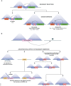
Figure 1. CTCF and cohesins build chromatin architecture
(A) Model of Topologically Associating Domains (TAD). TADs are regions of strong self‐contact. Promoter–enhancer interactions inside the domains are favoured while contacts with promoters and enhancers in adjacent domains are restrained. This is believed to help establish a functional organization of the genome. (B) Hi‐C profile illustrating TAD organization at an example locus in Neural Progenitor cells (data from Bonev et al, 2017). Increasing colour strength denotes enhanced interaction frequency. This in turn, can be interpreted as increased physical proximity in the three‐dimensional space of the cell nucleus. Triangles of Hi‐C signal reveal domains of enhanced interaction frequencies (TADs). Dots in the matrix (corner peaks) correspond to loops and reveal interactions between relatively short genomic intervals (here sub‐TAD boundaries). At some loci, TAD boundaries interact heavily with the entire TAD, which manifests itself as thin stripes of increased interaction frequency. (C) Loop extrusion model. Upon loading, cohesins (yellow ring) start translocating on chromatin (arrows) and their movement is accompanied by extrusion of an ever‐growing loop. Cohesins pass CTCF proteins bound to a motif which does not face them. Loop extrusion stops when cohesins encounter CTCF bound to a motif that is facing them (thick black arrow). (D) Model explaining the formation of architectural stripes. At genomic locations where cohesin loading occurs in the proximity of CTCF‐binding sites, including at active enhancers (green rectangle), CTCF bound to a motif oriented in a forward direction (en face) with respect to the loaded cohesin blocks loop extrusion immediately after loading. Loop extrusion proceeds fuelled by cohesin activity on the other side of the complex and allows the elements in the entire domain including promoters (red rectangle) to be “presented” to the fixed anchor overlapping the active enhancer (green rectangle). Depicted here is a single cohesin ring, it is unclear whether one or two cohesin rings extrude loops.
Figure 2
Figure 2. Genome engineering reveals locus‐specific transcriptional and architectural consequences of TAD boundary deletion and insertion
(A) TAD or a sub‐TAD boundary deletion may lead to no overt alteration of chromatin architecture as seen at the Firre locus (Barutcu et al, 2018), or to TAD and subTAD merging accompanied by either modest (Sox9/Kcjn2; Despang et al, 2019) or considerable transcriptional changes (e.g., loss of insulated neighbourhoods and oncogene activation; Hnisz et al, 2016), or aberrant activation of genes as, for example in the vicinity of otherwise insulated globin genes (Hanssen et al, 2017). (B) Ectopic insertion of a boundary element may lead to no change in the architecture of the recipient locus (Barutcu et al, 2018). When considering other CBS, boundaries can still be formed despite the deletion of the CBS. Depending on whether the ectopic boundary is inserted far or close to a Nipbl cohesin loader binding site, the boundary may form stripes (Redolfi et al, 2019). The contribution of distinct elements making up the boundary depends on the intrinsic features of the target locus. At one location, a boundary composed of a CBS site and a housekeeping gene promoter depends on both elements, while at another location CTCF appears less crucial for boundary formation (Zhang et al, 2020).
Figure 3
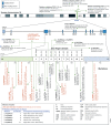
Figure 3. Mutations in CTCF related to neurological syndromes
Multiple mutations including deletions have been reported for CTCF. These genetic perturbations are linked to numerous neurological manifestations. Genetic variants impacting CTCF binding sites associate with several disorders including neurological diseases. The predicted impact of the mutations in ZFs of CTCF on its 3D protein structure and the inferred possible effects on CTCF binding to chromatin.

References

    1. Aitken SJ, Ibarra‐Soria X, Kentepozidou E, Flicek P, Feig C, Marioni JC, Odom DT (2018) CTCF maintains regulatory homeostasis of cancer pathways. Genome Biol 19: 106 - PMC - PubMed
    1. Akhtar W, de Jong J, Pindyurin AV, Pagie L, Meuleman W, de Ridder J, Berns A, Wessels LFA, van Lohuizen M, van Steensel B (2013) Chromatin position effects assayed by thousands of reporters integrated in parallel. Cell 154: 914–927 - PubMed
    1. Alexander JM, Guan J, Li B, Maliskova L, Song M, Shen Y, Huang B, Lomvardas S, Weiner OD (2019) Live‐cell imaging reveals enhancer‐dependent Sox2 transcription in the absence of enhancer proximity. elife 8: e41769 - PMC - PubMed
    1. Alipour E, Marko JF (2012) Self‐organization of domain structures by DNA‐loop‐extruding enzymes. Nucleic Acids Res 40: 11202–11212 - PMC - PubMed
    1. Aljahani A, Hua P, Karpinska MA, Quililan K, Davies JOJ, Oudelaar AM (2022) Analysis of sub‐kilobase chromatin topology reveals nano‐scale regulatory interactions with variable dependence on cohesin and CTCF. Nat Commun 13: 2139 - PMC - PubMed

Publication types