Review
. 2022 Oct 1;323(4):R375-R384.
doi: 10.1152/ajpregu.00151.2022. Epub 2022 Aug 22.

Neural control of the spleen as an effector of immune responses to inflammation: mechanisms and treatments

Affiliations
Review

Neural control of the spleen as an effector of immune responses to inflammation: mechanisms and treatments

Clarissa M D Mota et al. Am J Physiol Regul Integr Comp Physiol. .

Abstract

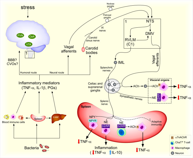
Immune system responses are a vital defense mechanism against pathogens. Inflammatory mediators finely regulate complex inflammatory responses from initiation to resolution. However, in certain conditions, the inflammation is initiated and amplified, but not resolved. Understanding the biological mechanisms underlying the regulation of the immune response is critical for developing therapeutic alternatives, including pharmaceuticals and bioelectronic tools. The spleen is an important immune effector organ since it orchestrates innate and adaptive immune responses such as pathogen clearance, cytokine production, and differentiation of cells, therefore playing a modulatory role that balances pro- and anti-inflammatory responses. However, modulation of splenic immune activity is a largely unexplored potential therapeutic tool that could be used for the treatment of inflammatory and life-threatening conditions. This review discusses some of the mechanisms controlling neuroimmune communication and the brain-spleen axis.

Keywords: autonomic nervous system; brain; inflammation; inflammatory reflex; neuroimmune axis.


Conflict of interest statement

No conflicts of interest, financial or otherwise, are declared by the authors.

Figures

Figure 1.
Figure 1.
Neural control of the spleen during endotoxemia. Bacterial antigens elicit a systemic inflammatory response. Cells and humoral factors emigrate from the spleen to the circulation and other organs. Inflammatory factors activate brain-mediated responses potentially via humoral (blood-brain-barrier, BBB, and circumventricular organs, CVOs) and neural routes (vagal and carotid sinus nerve afferents). Corticotropin-releasing hormone (CRH) neurons in central amygdala (CeA) and paraventricular nucleus (PVN) modulate adaptive immune responses to stress while ventral tegmental area (VTA) neurons modulate innate immune responses to bacteria. Peripheral afferents from the IX and X cranial nerves synapse with second-order neurons in the nucleus tractus solitarius (NTS), which can activate C1 neurons in the rostral ventrolateral medulla (RVLM) and neurons in the dorsal motor nucleus of the vagus (DMV). RVLM neurons synapse with preganglionic neurons in the intermediolateral column of the spinal cord (IML), including those giving rise to axons that form the splanchnic nerves. Axons from sympathetic ganglion cells in the celiac and suprarenal ganglia form the sympathetic splenic nerve, which nerve terminals release norepinephrine (NE) and neuropeptide Y (NPY). Choline acetyltransferase (ChAT)-expressing T-cells release acetylcholine (ACh) in response to β2-adrenergic receptor (AR) activation, leading to activation of α7 nicotinic receptor (α7nAChR) in macrophages. α7nAChR signaling blocks TNF-α release, thus reducing inflammation. β2-AR and NPY receptors (NPY-R) activation in immune cells reduces TNF-α production and β2-AR activation increases IL-10 release during inflammation. Afferent vagus nerve stimulation induces anti-inflammatory effects via increasing splenic nerve activity. Vagal efferent fibers send projections to sympathetic ganglia and viscera that, when stimulated, reduce inflammatory responses via β2-AR and α7nAChR. Dashed lines represent controversial or yet to be clarified pathways.

References

    1. Lewis SM, Williams A, Eisenbarth SC. Structure and function of the immune system in the spleen. Sci Immunol 4: eaau6085, 2019. doi:10.1126/sciimmunol.aau6085. - DOI - PMC - PubMed
    1. Cesta MF. Normal structure, function, and histology of the spleen. Toxicol Pathol 34: 455–465, 2006. doi:10.1080/01926230600867743. - DOI - PubMed
    1. Mebius RE, Kraal G. Structure and function of the spleen. Nat Rev Immunol 5: 606–616, 2005. doi:10.1038/nri1669. - DOI - PubMed
    1. Nance DM, Burns J. Innervation of the spleen in the rat: evidence for absence of afferent innervation. Brain Behav Immun 3: 281–290, 1989. doi:10.1016/0889-1591(89)90028-7. - DOI - PubMed
    1. Felten DL, Felten SY, Carlson SL, Olschowka JA, Livnat S. Noradrenergic and peptidergic innervation of lymphoid tissue. J Immunol 135, Suppl 2: 755s–765s, 1985. doi:10.1016/S0145-305X(06)80005-4. - DOI - PubMed

Publication types

LinkOut - more resources