. 2022 Sep 9;50(16):9505-9520.
doi: 10.1093/nar/gkac692.

Cryo-EM structure of the Smc5/6 holo-complex

Affiliations

Cryo-EM structure of the Smc5/6 holo-complex

Stephen T Hallett et al. Nucleic Acids Res. .

Abstract

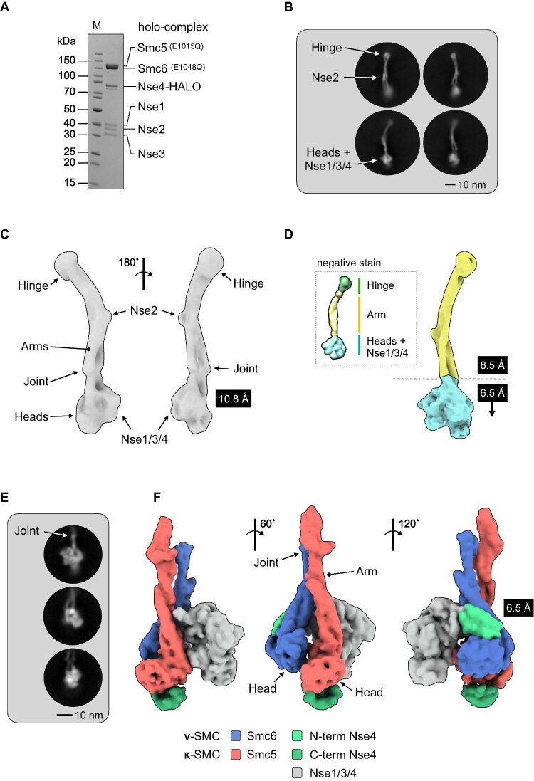
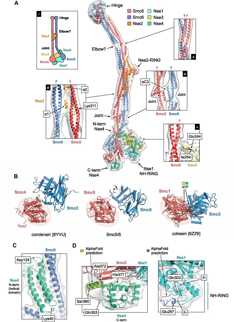
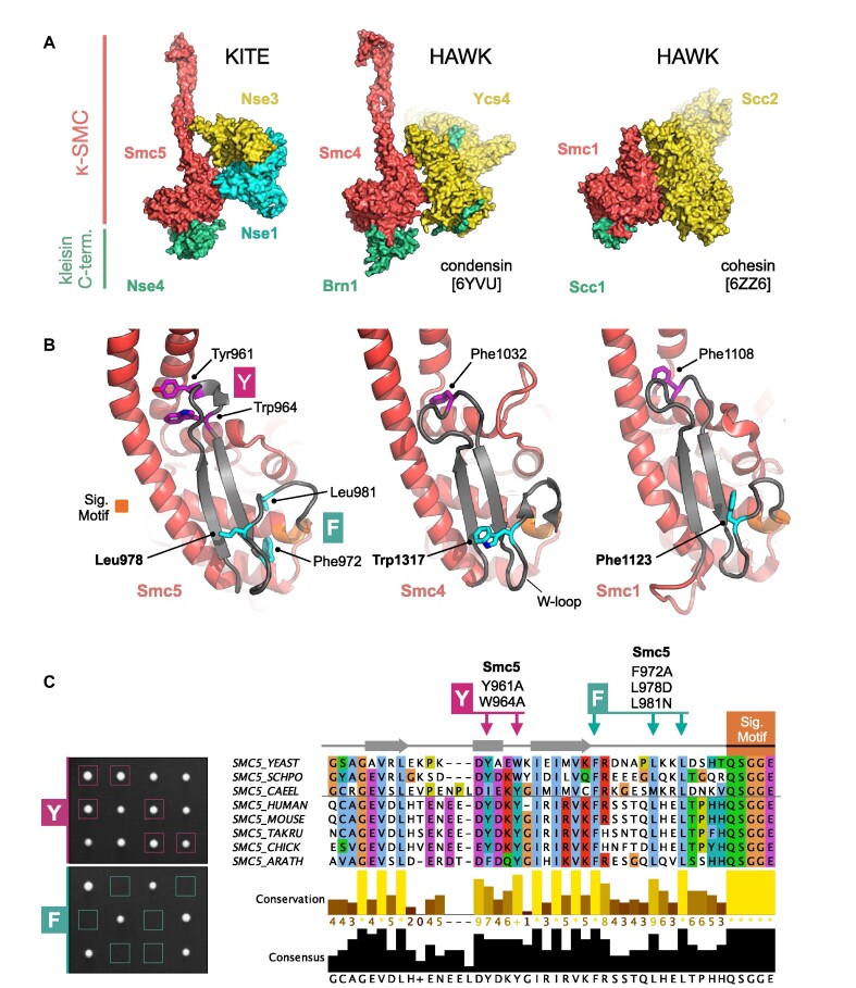
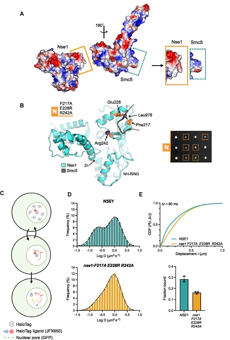
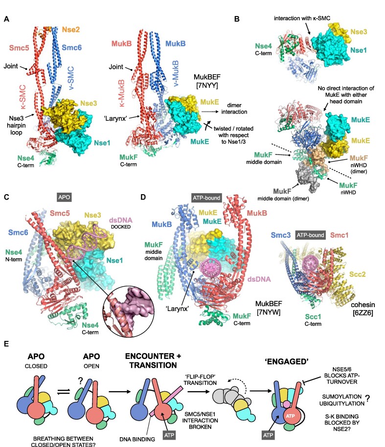
The Smc5/6 complex plays an essential role in the resolution of recombination intermediates formed during mitosis or meiosis, or as a result of the cellular response to replication stress. It also functions as a restriction factor preventing viral replication. Here, we report the cryogenic EM (cryo-EM) structure of the six-subunit budding yeast Smc5/6 holo-complex, reconstituted from recombinant proteins expressed in insect cells - providing both an architectural overview of the entire complex and an understanding of how the Nse1/3/4 subcomplex binds to the hetero-dimeric SMC protein core. In addition, we demonstrate that a region within the head domain of Smc5, equivalent to the 'W-loop' of Smc4 or 'F-loop' of Smc1, mediates an important interaction with Nse1. Notably, mutations that alter the surface-charge profile of the region of Nse1 which accepts the Smc5-loop, lead to a slow-growth phenotype and a global reduction in the chromatin-associated fraction of the Smc5/6 complex, as judged by single molecule localisation microscopy experiments in live yeast. Moreover, when taken together, our data indicates functional equivalence between the structurally unrelated KITE and HAWK accessory subunits associated with SMC complexes.


Figures

Figure 1.
Figure 1.
Cryo-EM of the budding yeast Smc5/6 complex. (A) Representative SDS-PAGE gel for the purified Smc5/6 holo-complex. (B) Representative 2D class averages (side views). Conformational flexibility leads to blurring of density at either the head (top) or hinge-end (bottom) of the complex. (C) Initial 3D map from cryo-EM at a resolution of 10.8 Å. (D) Maps from focussed refinement at 8.5Å and 6.5 Å for the indicated segments of the Smc5/6 holo-complex (d, inset) model obtained by uranyl acetate negative stain electron microscopy for comparison (25). (E) Representative 2D class averages (side views) and (F) resultant 3D map at 6.5 Å for the ‘head’-end of the complex. The cryo-EM map has been segmented and coloured with respect to its assigned component (see associated key for additional detail).
Figure 2.
Figure 2.
A pseudo-atomic model for the Smc5/6 holo-complex. (A) Overview of the pseudo-atomic model. (A, inset i) Schematic showing the overall architecture of the Smc5/6 complex and selected molecular features. (A, inset ii) Expanded view of the ‘Elbow’, highlighting the crossover of the coiled-coil ‘arms’ of Smc5 and Smc6 at this point. (A, inset iii) The first alpha-helix of Nse2 (α1) is situated between the two arms of the complex. The position of Lys311, a known site of auto-SUMOylation is also indicated (A, inset iv) Expanded view of the interface between the ‘Joint’ features of Smc5 and Smc6, involving the two αC3 helices. (A, inset v) A short beta-hairpin (amino acids Glu244-Ile254) protruding from Nse3 is in close proximity to the arm of Smc5. For each inset, the directionality of the ascending helix (head to hinge) is indicated by a blue or red arrow, for Smc5 and Smc6 respectively. (B) Comparison of the relative head domain positions in the cryo-EM structures of budding yeast condensin (PDB: 6YVU), Smc5/6 (this manuscript) and cohesin (PDB: 6ZZ6); in each, using the head of the κ-SMC as a fixed reference point. (C) Expanded view for the N-terminal helical domain of Nse4 (aa Lys40-Asp124) bound to the ‘arm’ of Smc6. (D, left) AlphaFold predicts the presence of an additional helical element (aa Ser360-Ala372) in the C-terminal domain of Nse4. (D, right). AlphaFold predicts a budding yeast-specific loop insertion in the NH-RING of Nse1 (aa Glu287-Gln303). Where shown, sections of density from the composite cryo-EM map are represented by a semi-transparent molecular surface, shaded in grey. Please also see associated key for additional detail.
Figure 3.
Figure 3.
KITES and HAWKS share a common interaction interface involving the κ-SMC ‘W-loop’. (A) Side-by-side visualisation of the κ-SMC head domain from Smc5/6 (left), condensin (middle) and cohesin (right) in complex with their respective kleisin C-terminal domain; Nse4, Brn1 and Scc1. In each case, the interacting partner, whether KITE or HAWK, makes a similar set of interactions with the head domain of the κ-SMC. (B) Expanded view, showing secondary structure molecular cartoons for each κ-SMC head domain, highlighting the position of conserved amino acids within the ‘W-loop’ or equivalent (stick representation, carbon atoms coloured cyan) plus aromatic residues within the preceding sequence (stick representation, carbon atoms coloured magenta). The ABC-signature motif is additionally highlighted in orange. (C, left) Tetrad dissections. Spores derived from diploid S. cerevisiae strains carrying both wild-type allele and indicated mutant allele plus associated NAT-selectable marker (natMX6). Genotypes were confirmed by replica plating of spores on selective media (not shown). (C, right) Multiple sequence alignment, across selected species, showing conservation and consensus of amino acids within the W-loop and preceding region of Smc5 (produced using Jalview 2 with Clustal X colour scheme; (73)). Sets of compound mutations introduced into budding yeast: Smc5-Y = Y961A, W964A; Smc5-F = F972A, L978D, L981N. Please also see associated key for additional detail.
Figure 4.
Figure 4.
Examining the cellular effects of breaking the Nse1 / Smc5-loop interface. (A) Molecular surface representations for Nse1 and the head domain of Smc5, coloured by electrostatic potential (APBS plugin, PyMOL). The surface of Nse1 and the conserved loop extending from Smc5 display a high degree of charge complimentary (visualisation aided by rotation of the head domain through 180°. The interacting regions, as bounded by the drawn rectangles, are shown in an expanded view on the right. (B, left) Molecular secondary structure cartoon representation, showing the relative locations of the amino acids mutated within the Nse1/Smc5-loop interface: Nse1-N = F217A, E228R, R242A (b, right) Tetrad dissection. Spores derived from diploid S. cerevisiae strains carrying both wild-type and indicated mutant allele plus associated NAT-selectable marker (natMX6). Genotypes were confirmed by replica plating of spores on selective media (not shown). (C) Schematic of our single particle tracking approach. Yeast endogenously expressing Nse4 fused to a C-terminal HaloTag are labelled with JFX650 dye. Fluorescence from individual dye molecules is then used to calculate trajectories that represent the diffusion behaviour of the Smc5/6 complex containing the HaloTag fused Nse4 subunit. (D) Diffusion coefficient frequency histograms calculated using pooled data (three independent experimental repeats) in either the NSE1 or nse1-F217A E228R R242A genetic backgrounds. (E, top), Cumulative distribution frequency plot of pooled single-molecule displacements. (E, bottom) ‘Fraction bound’ values determined from fitting of experimental data with kinetic models available within Spot-On. Filled circles represent the value determined from each technical repeat, with the height of the bar corresponding to the mean. Error bars represent one standard deviation. Please also see associated keys for additional details.
Figure 5.
Figure 5.
Exploration of conformational changes likely to accompany binding of dsDNA/ATP. (A) Comparison of the apo-states for Smc5/6 and the related MukBEF complex (PDB: 7NYY). To aid visualisation, only subunits with direct equivalence across both complexes are shown, and for clarity molecular surfaces instead of secondary structure cartoons are shown for the respective KITE proteins. A direct contact between the ‘joint’ features of Smc5 and Smc6 serves to generates a fully ‘closed’ conformation similar to that reported for MukBEF (64) but without a reinforcing secondary ‘larynx’ interface. Furthermore, the KITE homodimer formed by MukE sits, and is held, in a different position when compared to the KITE heterodimer of Nse1/Nse3, largely as a result of its more elaborate interaction with its dimeric kleisin partner MukF. (B) In the apo-state, the MukE heterodimer makes no direct interaction with the head domain of either MukB protein, instead making a series of interactions with the domain-swapped N-terminal winged-helix domain (nWHD) of MukF that anchor it in place. (C) Simple superposition of a DNA duplex, taken from the docking pose reported for the human NSE1/3 heterodimer (65) onto our apo-state structure, indicates that without accompanying conformational changes extension of the trajectory for the bound DNA would generate steric clashes with the arm of Smc5 (inset, DNA now shown in surface representation). (D) DNA/ATP-bound forms of MukBEF (left) and cohesin (inset, right), providing side-by-side comparisons and a visualisation aid of the expected fully ‘engaged’ conformation of SMC-complexes. (E) A speculative model for how the Smc5/6 complex might bind to and engage with dsDNA. We propose that the apo-state can ‘breathe’ between a fully closed conformation and a more open state, which allows / facilitates binding of dsDNA to the positively charged surface / groove created at the interface of Nse1/Nse3, to generate an intermediary ‘encounter’ complex. This, along with concomitant binding of ATP to the head domain of Smc5, serves to break the Smc5/Nse1 interaction allowing a ‘flip-flop’-type transition to the anticipated fully ‘engaged’ state. It is not clear how, or indeed if, ubiquitylation, SUMOylation or other post-translational modification affects either conformation or ATPase activity. It is also not known if the presence of Nse2, acts to block binding or transition of bound dsDNA into the S-K ring (SMC-kleisin) compartment. Binding of the Nse5/6 heterodimer blocks the ability of Smc5/6 to turn over ATP (22,25), but it is not fully known what effect this has on the overall conformation at the head-end of the complex.

References

    1. Cutts E.E., Vannini A.. Condensin complexes: understanding loop extrusion one conformational change at a time. Biochem. Soc. Trans. 2020; 48:2089–2100. - PMC - PubMed
    1. Datta S., Lecomte L., Haering C.H.. Structural insights into DNA loop extrusion by SMC protein complexes. Curr. Opin. Struct. Biol. 2020; 65:102–109. - PMC - PubMed
    1. Hassler M., Shaltiel I.A., Haering C.H.. Towards a unified model of SMC complex function. Curr. Biol. 2018; 28:R1266–R1281. - PMC - PubMed
    1. Matityahu A., Onn I.. Hit the brakes - a new perspective on the loop extrusion mechanism of cohesin and other SMC complexes. J. Cell Sci. 2021; 134:jcs247577. - PubMed
    1. Palecek J.J. SMC5/6: multifunctional player in replication. Genes (Basel). 2018; 10:7. - PMC - PubMed

Publication types

MeSH terms

Substances