Editorial
. 2022 Dec 1;28(23):5121-5135.
doi: 10.1158/1078-0432.CCR-22-1493.

Activation of Adaptive and Innate Immune Cells via Localized IL2 Cytokine Factories Eradicates Mesothelioma Tumors

Affiliations
Editorial

Activation of Adaptive and Innate Immune Cells via Localized IL2 Cytokine Factories Eradicates Mesothelioma Tumors

Amanda M Nash et al. Clin Cancer Res. .

Abstract

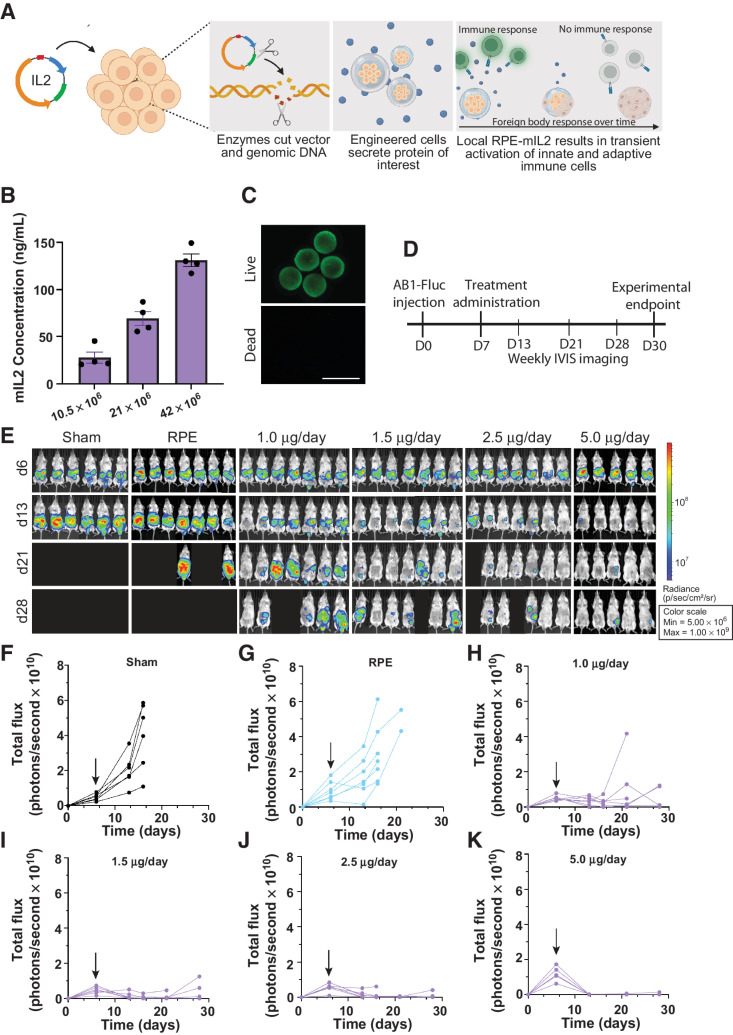
Purpose: IL2 immunotherapy has the potential to elicit immune-mediated tumor lysis via activation of effector immune cells, but clinical utility is limited due to pharmacokinetic challenges as well as vascular leak syndrome and other life-threatening toxicities experienced by patients. We developed a safe and clinically translatable localized IL2 delivery system to boost the potency of therapy while minimizing systemic cytokine exposure.

Experimental design: We evaluated the therapeutic efficacy of IL2 cytokine factories in a mouse model of malignant mesothelioma. Changes in immune populations were analyzed using time-of-flight mass cytometry (CyTOF), and the safety and translatability of the platform were evaluated using complete blood counts and serum chemistry analysis.

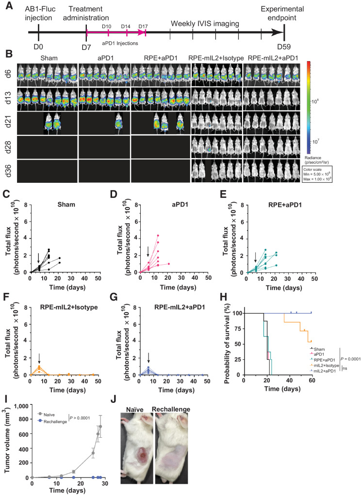
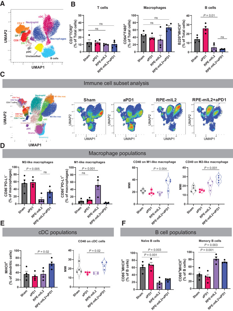
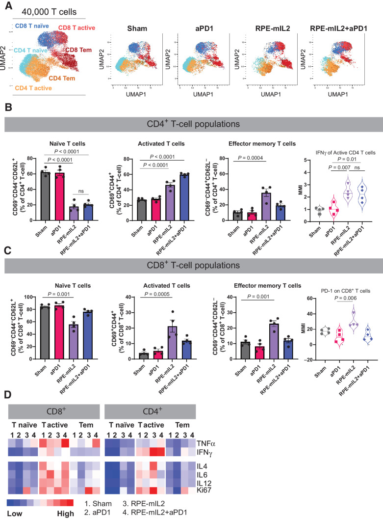
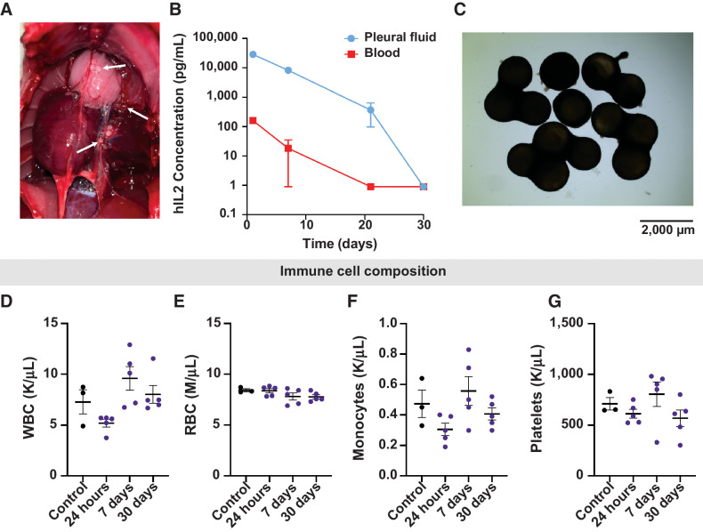
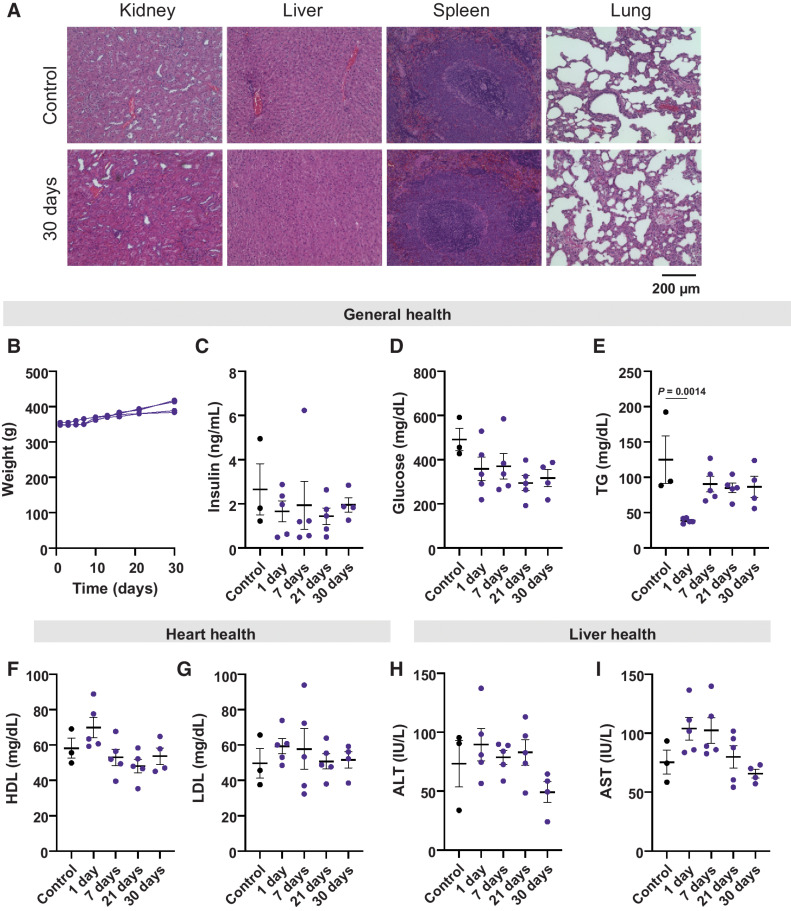
Results: IL2 cytokine factories enabled 150× higher IL2 concentrations in the local compartment with limited leakage into the systemic circulation. AB1 tumor burden was reduced by 80% after 1 week of monotherapy treatment, and 7 of 7 of animals exhibited tumor eradication without recurrence when IL2 cytokine factories were combined with anti-programmed cell death protein 1 (aPD1). Furthermore, CyTOF analysis showed an increase in CD69+CD44+ and CD69-CD44+CD62L- T cells, reduction of CD86-PD-L1- M2-like macrophages, and a corresponding increase in CD86+PD-L1+ M1-like macrophages and MHC-II+ dendritic cells after treatment. Finally, blood chemistry ranges in rodents demonstrated the safety of cytokine factory treatment and reinforced its potential for clinical use.

Conclusions: IL2 cytokine factories led to the eradication of aggressive mouse malignant mesothelioma tumors and protection from tumor recurrence, and increased the therapeutic efficacy of aPD1 checkpoint therapy. This study provides support for the clinical evaluation of this IL2-based delivery system. See related commentary by Palanki et al., p. 5010.


Figures

Figure 1. Dose response of RPE-mIL2 in a peritoneal model of malignant mesothelioma. A, Schematic demonstrating the development of RPE-mIL2 cells and encapsulation in hydrogel spheres. B, ELISA measurements of mIL2 in supernatant collected from capsules after 24 hours of in vitro culture C, Representative live/dead image of RPE-mIL2 cells encapsulated at 42e6 cells/mL in alginate. D, Schematic illustrating the experimental timeline for tumor establishment, treatment, and IVIS imaging. E, Luminescent images tracking AB1-FLuc tumor burden over time beginning at day 6 postinjection, and weekly until day 28 post injection. Subsequent to stratification, the image of each mouse was individually cropped and stitched to create a collage of each treatment group. F–K, Quantification of tumor burden for each treatment group (n = 5–7) represented by total flux (photons/second) plotted over time. Black arrows indicate the day of treatment administration.
Figure 1.
Dose response of RPE-mIL2 in a peritoneal model of malignant mesothelioma. A, Schematic demonstrating the development of RPE-mIL2 cells and encapsulation in hydrogel spheres. B, ELISA measurements of mIL2 in supernatant collected from capsules after 24 hours of in vitro culture. C, Representative live/dead image of RPE-mIL2 cells encapsulated at 42 × 106 cells/mL in alginate. D, Schematic illustrating the experimental timeline for tumor establishment, treatment, and IVIS imaging. E, Luminescent images tracking AB1-FLuc tumor burden over time beginning at day 6 post injection, and weekly until day 28 post injection. Subsequent to stratification, the image of each mouse was individually cropped and stitched to create a collage of each treatment group. F–K, Quantification of tumor burden for each treatment group (n = 5–7) represented by total flux (photons/second) plotted over time. Black arrows indicate the day of treatment administration.
Figure 2. RPE-mIL2 improves therapeutic efficacy of aPD1. A, Schematic of the experimental timeline for tumor establishment, treatment administration, and IVIS imaging. B, Luminescent images tracking tumor burden over time. Subsequent to stratification, the image of each mouse was individually cropped and stitched to create a collage of each treatment group. C–G, Quantification of tumor burden for each treatment group (n = 7–8) represented by total flux (photons/second) plotted over time. Black arrows indicate the day of treatment administration. H, Survival curves plotted as percent survival over time beginning after tumor injection (n = 7–8). P value was determined by a comparison of survival curves by the log-rank (Mantel–Cox) test (ns = not significant). I, plot of subcutaneous tumor volume over time in naïve mice compared with RPE-mIL2+aPD1 treated mice. P values were acquired using one way ANOVA with Holm–Sidak method for multiple comparisons. J, Representative macroscopic images of the left flank 28 days post subcutaneous tumor injection. (Left; naïve, Right; RPE-mIL2+aPD1 treated).
Figure 2.
RPE-mIL2 improves therapeutic efficacy of aPD1. A, Schematic of the experimental timeline for tumor establishment, treatment administration, and IVIS imaging. B, Luminescent images tracking tumor burden over time. Subsequent to stratification, the image of each mouse was individually cropped and stitched to create a collage of each treatment group. C–G, Quantification of tumor burden for each treatment group (n = 7–8) represented by total flux (photons/second) plotted over time. Black arrows indicate the day of treatment administration. H, Survival curves plotted as percent survival over time beginning after tumor injection (n = 7–8). P value was determined by a comparison of survival curves by the log-rank (Mantel–Cox) test (ns = not significant). I, plot of subcutaneous tumor volume over time in naïve mice compared with RPE-mIL2+aPD1–treated mice. P values were acquired using one-way ANOVA with Holm–Sidak method for multiple comparisons. J, Representative macroscopic images of the left flank 28 days post subcutaneous tumor injection. (Left; naïve, Right; RPE-mIL2+aPD1 treated).
Figure 3. Alteration of immune composition after RPE-mIL2 or RPE-mIL2+aPD1 therapy. A, Immune atlas map. We performed CyTOF with single cell suspension obtained from peritoneal lavage fluid. The Uniform Manifold Approximation and Projection (UMAP) was applied for dimensional reduction with 1,000,000 cells (40,000/each experiment × 4 mice/group × 4 groups). B, Comparison of T cells, macrophages, and B cells across treatment groups (n = 4 per group). C, UMAP of specific immune cell subsets. RPE-mIL2 treatment and combination of aPD1 therapy with mIL2 led to dramatic changes in lymphocytes and myeloid cell compositions. D, Comparison of M1-like and M2-like macrophages across treatment groups. Expression of CD40 among M1-like or M2-like macrophages across treatment groups. E, Comparison of cDC cells across treatment groups. Expression of CD40 among cDC cells across treatment groups. F, Comparison of naïve and memory B cells across treatment groups. P values were acquired using one way ANOVA with Holm–Sidak method for multiple comparisons, ns = not significant.
Figure 3.
Alteration of immune composition after RPE-mIL2 or RPE-mIL2+aPD1 therapy. A, Immune atlas map. We performed CyTOF with single cell suspension obtained from peritoneal lavage fluid. The Uniform Manifold Approximation and Projection (UMAP) was applied for dimensional reduction with 1,000,000 cells (40,000/each experiment × 4 mice/group × 4 groups). B, Comparison of T cells, macrophages, and B cells across treatment groups (n = 4 per group). C, UMAP of specific immune cell subsets. RPE-mIL2 treatment and combination of aPD1 therapy with mIL2 led to dramatic changes in lymphocytes and myeloid cell compositions. D, Comparison of M1-like and M2-like macrophages across treatment groups. Expression of CD40 among M1-like or M2-like macrophages across treatment groups. E, Comparison of cDC cells across treatment groups. Expression of CD40 among cDC cells across treatment groups. F, Comparison of naïve and memory B cells across treatment groups. P values were acquired using one-way ANOVA with Holm–Sidak method for multiple comparisons, ns = not significant.
Figure 4. Alteration of T-cell subsets after RPE-mIL2 or RPE-mIL2+aPD1 therapy. A, UMAP of specific T-cell subsets analyzed in this study. B, Comparison of naïve, activated, or effector memory CD4+ T cells across treatment groups. Expression of IFNγ among activated CD4+ T cells across treatment groups. C, Comparison of naïve, activated, or effector memory CD8+ T cells across treatment groups. Expression of PD-1 among activated CD8+ T cells across treatment groups (n = 4 per group). D, Heatmap of relative TNFα, IFNγ, IL4, IL6, IL12 and Ki67 expression across each group. P values were acquired using one way ANOVA with Holm–Sidak method for multiple comparisons, ns = not significant.
Figure 4.
Alteration of T-cell subsets after RPE-mIL2 or RPE-mIL2+aPD1 therapy. A, UMAP of specific T-cell subsets analyzed in this study. B, Comparison of naïve, activated, or effector memory CD4+ T cells across treatment groups. Expression of IFNγ among activated CD4+ T cells across treatment groups. C, Comparison of naïve, activated, or effector memory CD8+ T cells across treatment groups. Expression of PD-1 among activated CD8+ T cells across treatment groups (n = 4 per group). D, Heatmap of relative TNFα, IFNγ, IL4, IL6, IL12, and Ki67 expression across each group. P values were acquired using one-way ANOVA with Holm–Sidak method for multiple comparisons, ns = not significant.
Figure 5. Evaluation of human IL2 kinetics in the rat pleura. A, Macroscopic image of the pleural cavity 24 hours post-implant. White arrows indicate RPE-hIL2. B, ELISA measurements of hIL2 concentration in the pleural cavity and blood at 24 hours, 7 days, 21 days, and 30 days post implant. C, Bright field images of capsules retrieved from the pleural cavity 30 days post implant. Image acquired at 2x magnification. D-G, plots of white blood cells, red blood cell, monocyte, and platelet concentration derived from complete blood count analysis at 24 hours, 7 days, and 30 days post implant (n = 5 per group). Values are compared to untreated controls. Differences in cell concentration were not significant across all groups. P values were acquired using one way ANOVA with Holm–Sidak method for multiple comparisons.
Figure 5.
Evaluation of human IL2 kinetics in the rat pleura. A, Macroscopic image of the pleural cavity 24 hours post implant. White arrows indicate RPE-hIL2. B, ELISA measurements of hIL2 concentration in the pleural cavity and blood at 24 hours, 7 days, 21 days, and 30 days post implant. C, Bright field images of capsules retrieved from the pleural cavity 30 days post implant. Image acquired at 2× magnification. D-G, Plots of white blood cell, red blood cell, monocyte, and platelet concentration derived from complete blood count analysis at 24 hours, 7 days, and 30 days post implant (n = 5 per group). Values are compared with untreated controls. Differences in cell concentration were not significant across all groups. P values were acquired using one-way ANOVA with Holm–Sidak method for multiple comparisons.
Figure 6. Toxicological analysis of hIL2 in the rat pleura over the span of 30 days. A, Representative images of H&E-stained sections of the kidney, liver, spleen, and lungs of untreated control rats compared with rats sacrificed at 30 days post capsule implant (n = 5). B, Plot tracking the weight of individual rats over the course of treatment. C and D, evaluation of general health through changes in insulin and glucose for each time point. Values are compared to untreated controls. Differences in values were not significant across all groups. P values were acquired using one way ANOVA with Holm–Sidak method for multiple comparisons. E, Evaluation of general health through changes in Triglycerides for each time point. F and G, evaluation of heart health through changes in HDL and LDL levels over time. H and I, Evaluation of liver health through changes in ATL and AST levels over time. All values are compared to untreated controls. Differences in values were not significant across all groups. P values were acquired using one way ANOVA with Holm–Sidak method for multiple comparisons.
Figure 6.
Toxicological analysis of hIL2 in the rat pleura over the span of 30 days. A, Representative images of H&E-stained sections of the kidney, liver, spleen, and lungs of untreated control rats compared with rats sacrificed at 30 days post capsule implant (n = 5). B, Plot tracking the weight of individual rats over the course of treatment. C and D, Evaluation of general health through changes in insulin and glucose for each time point. Values are compared with untreated controls. Differences in values were not significant across all groups. P values were acquired using one-way ANOVA with Holm–Sidak method for multiple comparisons. E, Evaluation of general health through changes in triglycerides for each time point. F and G, Evaluation of heart health through changes in HDL and LDL levels over time. H and I, Evaluation of liver health through changes in ALT and AST levels over time. All values are compared with untreated controls. Differences in values were not significant across all groups. P values were acquired using one-way ANOVA with Holm–Sidak method for multiple comparisons.

Comment in

Similar articles

Cited by

References

    1. Nelson DB, Rice DC, Niu J, Atay S, Vaporciyan AA, Antonoff M, et al. . Long-term survival outcomes of cancer-directed surgery for malignant pleural mesothelioma: propensity score matching analysis. J Clin Oncol 2017;35:3354–62. - PubMed
    1. Meyerhoff RR, Yang CF, Speicher PJ, Gulack BC, Hartwig MG, D'Amico TA, et al. . Impact of mesothelioma histologic subtype on outcomes in the surveillance, epidemiology, and end results database. J Surg Res 2015;196:23–32. - PMC - PubMed
    1. Musk AW, Olsen N, Alfonso H, Reid A, Mina R, Franklin P, et al. . Predicting survival in malignant mesothelioma. Eur Respir J 2011;38:1420–4. - PubMed
    1. Linton A, Pavlakis N, O'Connell R, Soeberg M, Kao S, Clarke S, et al. . Factors associated with survival in a large series of patients with malignant pleural mesothelioma in New South Wales. Br J Cancer 2014;111:1860–9. - PMC - PubMed
    1. Taioli E, Wolf AS, Camacho-Rivera M, Kaufman A, Lee DS, Nicastri D, et al. . Determinants of survival in malignant pleural mesothelioma: a surveillance, epidemiology, and end results (SEER) Study of 14,228 patients. PLoS One 2015;10:e0145039. - PMC - PubMed

Publication types