. 2022 Dec 2;18(11):e872-e887.
doi: 10.4244/EIJ-E-22-00036.

Pre-procedural planning of coronary revascularization by cardiac computed tomography: An expert consensus document of the Society of Cardiovascular Computed Tomography

Affiliations

Pre-procedural planning of coronary revascularization by cardiac computed tomography: An expert consensus document of the Society of Cardiovascular Computed Tomography

Daniele Andreini et al. EuroIntervention. .

Abstract

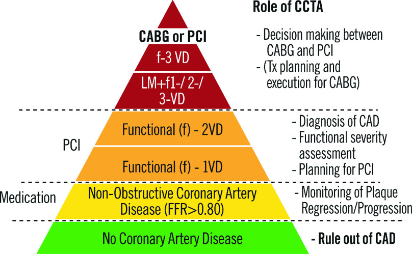
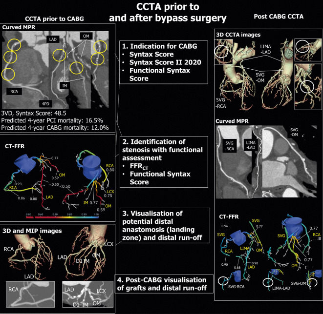
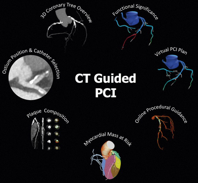
Coronary CT angiography (CCTA) demonstrated high diagnostic accuracy for detecting coronary artery disease (CAD) and a key role in the management of patients with low-to-intermediate pretest likelihood of CAD. However, the clinical information provided by this noninvasive method is still regarded insufficient in patients with diffuse and complex CAD and for planning percutaneous coronary intervention (PCI) and surgical revascularization procedures. On the other hand, technology advancements have recently shown to improve CCTA diagnostic accuracy in patients with diffuse and calcific stenoses. Moreover, stress CT myocardial perfusion imaging (CT-MPI) and fractional flow reserve derived from CCTA (CT-FFR) have been introduced in clinical practice as new tools for evaluating the functional relevance of coronary stenoses, with the possibility to overcome the main CCTA drawback, i.e. anatomical assessment only. The potential value of CCTA to plan and guide interventional procedures lies in the wide range of information it can provide: a) detailed evaluation of plaque extension, volume and composition; b) prediction of procedural success of CTO PCI using scores derived from CCTA; c) identification of coronary lesions requiring additional techniques (e.g., atherectomy and lithotripsy) to improve stent implantation success by assessing calcium score and calcific plaque distribution; d) assessment of CCTA-derived Syntax Score and Syntax Score II, which allows to select the mode of revascularization (PCI or CABG) in patients with complex and multivessel CAD. The aim of this Consensus Document is to review and discuss the available data supporting the role of CCTA, CT-FFR and stress CT-MPI in the preprocedural and possibly intraprocedural planning and guidance of myocardial revascularization interventions.


Figures

Figure 1
Figure 1. Image showing a co-registration of invasive coronary angiography (A), coronary CTA and straight MPR (panel B and C) with CTA cross sections (panel D), corresponding OCT cross sections and longitudinal OCT view (E).
Panel F shows the CT-FFR patient-specific model and panel G the result of the CT-FFR Planner. Invasive coronary angiography (A), coronary CT angiography (B) and straight MPR (C) showing a calcified fibro-atheroma with positive remodeling in the proximal mid LAD. The CT-FFR (panel F) reaches a value of 0.59 with a focal gradient at the level of the MLA (panel D and E mid). The OCT cross sections mirror the CTA images. The most distal CTA cross section exhibits a healthy landing zone for percutaneous stenting also visualized by OCT (E). Based on the CT-FFR model (F), an appropriate stent length and position is selected (dashed lines). In panel G the predicted functional result by the CT-FFR Planner mimicking PCI can be appreciated.
Figure 2
Figure 2. Stress CT-MPI for planning PCI in patients with known CAD.
A complete protocol of adenosine stress-rest CT-MPI was performed. In panels A-C coronary anatomy shows a critical LAD stenosis (panel A), occlusion of LCX (panel B) and patency of a previous stent in the RCA (panel C); the same scan demonstrates reversible perfusion deficit of the antero-septal wall and only partial reversible perfusion deficit of the posterolateral wall (panel D-G). Invasive coronary angiography confirms LCX occlusion and a critical stenosis of the LAD (panel H-L). The interventional cardiologist was able to plan the revascularization procedure and, more importantly, to decide with the Heart Team whether to treat LCX occlusion before bringing the patient to the cath lab.
Figure 3
Figure 3. CT-Rector Score Calculator.
Figure 4
Figure 4. KCCT Score: definition and scoring system.
Figure 5
Figure 5. Role of CCTA in different degrees of CAD.
Figure 6
Figure 6. CCTA prior to and after Bypass surgery.
Left side: CCTA before CABG. Upper panel: Curved multiplanar reconstructions of major epicardial vessels showing the lesions scored in the SYNTAX score. Mid panel: CT-FFR analysis. Lower panel: 3D Volume rendering and MIP reconstructions. Right side: CCTA after CABG. Upper panel: 3D Volume rendering images showing the three grafts on LAD, OM and RCA. Mid panel: Curved multiplanar reconstructions of the three grafts. Lower panel: CT-FFR of native arteries and grafts. LAD = left anterior descending artery; MIP = maximum intensity projection; OM = obtuse marginal branch; RCA = right coronary artery; 3D = three dimensional.
Figure 7
Figure 7. Prototype of coronary CT angiography catheterization laboratory viewer for online PCI guidance.
Software prototype allowing for visualization of the coronary anatomy and plaque components extracted from coronary CT angiography (CCTA) inside the catheterization laboratory. The central panel shows a three-dimensional (3D) reconstruction of a left coronary artery (red) with colored-coded visualization of the plaque components (white for calcium and green for non-calcified plaque). The inferior panel shows a straight MPR reconstruction with three markers (green, blue and red lines) related to 3D reconstruction. This tool allows for the measurement of lesion length for stent length selection. The top right panel shows a CCTA cross-sectional view of the left anterior descending artery displaying the lumen and vessel contours (yellow and orange lines). The dimension of the lumen and vessel, both mean diameter and area, are shown in the right mid-panel, and in the bottom right corner, the diameter function line in the y-axis and length in the x-axis. Software prototype QAngio Cath Lab from Medis Medical Imaging. (For interpretation of the references to color in this figure legend, the reader is referred to the Web version of this article.)
Figure 8
Figure 8. Main features of Cardiac CT-guided PCI.
CCTA-derived parameters for PCI planning and guidance. The figure shows the main features of a CT-guided PCI approach, including assessment of coronary ostium position for guiding catheter selection, 3-vessel overview of coronary tree for evaluation of calcium extent and distribution, plaque composition and quantification analysis, functional significance of stenosis and virtual PCI planning by CT-FFR, estimation of myocardial mass at risk and online procedural guidance.

Similar articles

Cited by

References

    1. Stein PD, Yaekoub AY, Matta F, Sostman HD. 64-slice CT for diagnosis of coronary artery disease: a systematic review. Am J Med. 2008;121(8):715–25. - PubMed
    1. Schroeder S, Achenbach S, Bengel F, et al. Working group nuclear cardiology and cardiac CT; European Society of cardiology; European Council of nuclear cardiology. Cardiac computed tomography: indications, applications, limitations, and training requirements: report of a writing group deployed by the working group nuclear cardiology and cardiac CT of the European Society of cardiology and the European Council of nuclear cardiology. Eur Heart J. 2008;29(4):531–56. - PubMed
    1. Budoff MJ, Dowe D, Jollis JG, et al. Diagnostic performance of 64-multidetector row coronary computed tomographic angiography for evaluation of coronary artery stenosis in individuals without known coronary artery disease: results from the prospective multicenter ACCURACY (Assessment by Coronary Computed Tomographic Angiography of Individuals Undergoing Invasive Coronary Angiography) trial. J Am Coll Cardiol. 2008;52(21):1724–32. - PubMed
    1. Meijboom WB, Meijs MF, Schuijf JD, et al. Diagnostic accuracy of 64-slice computed tomography coronary angiography: a prospective, multicenter, multivendor study. J Am Coll Cardiol. 2008;52(25):2135–44. - PubMed
    1. Marano R, De Cobelli F, Floriani I, et al, NIMISCAD Study Group. Italian multicenter, prospective study to evaluate the negative predictive value of 16- and 64-slice MDCT imaging in patients scheduled for coronary angiography (NIMISCAD-Non Invasive Multicenter Italian Study for Coronary Artery Disease). Eur Radiol. 2009;19(5):1114–23. - PubMed

MeSH terms