. 2022 Sep;227(7):2553-2567.
doi: 10.1007/s00429-022-02546-2. Epub 2022 Aug 22.

Multimodal tract-based MRI metrics outperform whole brain markers in determining cognitive impact of small vessel disease-related brain injury

Collaborators, Affiliations

Multimodal tract-based MRI metrics outperform whole brain markers in determining cognitive impact of small vessel disease-related brain injury

Alberto De Luca et al. Brain Struct Funct. 2022 Sep.

Abstract

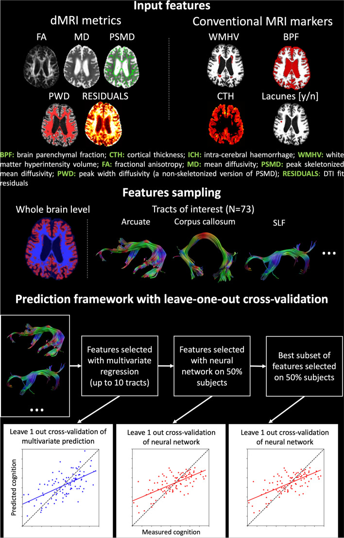
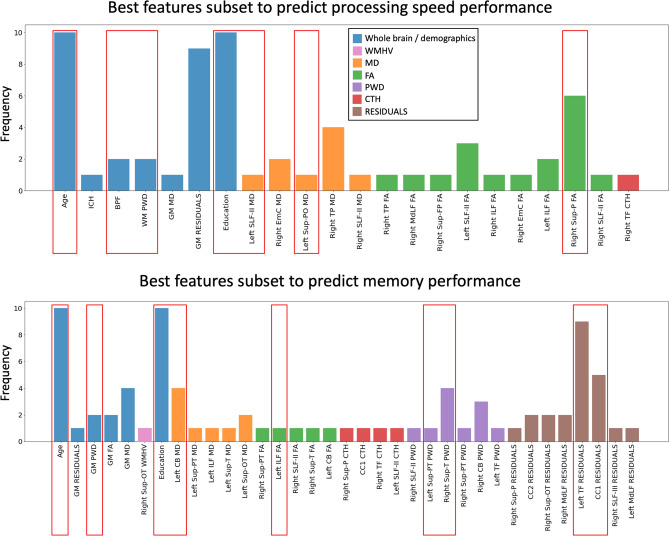
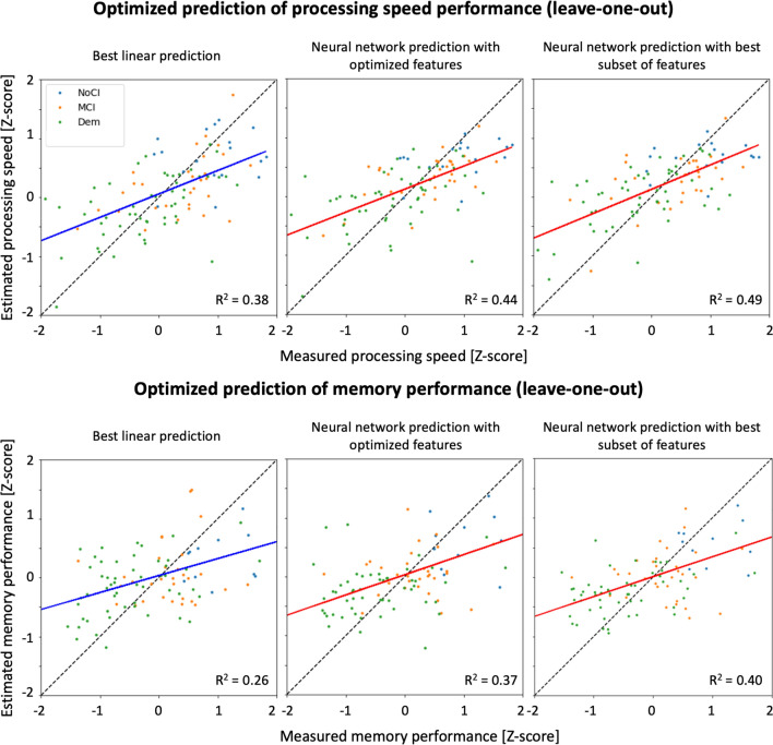
In cerebral small vessel disease (cSVD), whole brain MRI markers of cSVD-related brain injury explain limited variance to support individualized prediction. Here, we investigate whether considering abnormalities in brain tracts by integrating multimodal metrics from diffusion MRI (dMRI) and structural MRI (sMRI), can better capture cognitive performance in cSVD patients than established approaches based on whole brain markers. We selected 102 patients (73.7 ± 10.2 years old, 59 males) with MRI-visible SVD lesions and both sMRI and dMRI. Conventional linear models using demographics and established whole brain markers were used as benchmark of predicting individual cognitive scores. Multi-modal metrics of 73 major brain tracts were derived from dMRI and sMRI, and used together with established markers as input of a feed-forward artificial neural network (ANN) to predict individual cognitive scores. A feature selection strategy was implemented to reduce the risk of overfitting. Prediction was performed with leave-one-out cross-validation and evaluated with the R2 of the correlation between measured and predicted cognitive scores. Linear models predicted memory and processing speed with R2 = 0.26 and R2 = 0.38, respectively. With ANN, feature selection resulted in 13 tract-specific metrics and 5 whole brain markers for predicting processing speed, and 28 tract-specific metrics and 4 whole brain markers for predicting memory. Leave-one-out ANN prediction with the selected features achieved R2 = 0.49 and R2 = 0.40 for processing speed and memory, respectively. Our results show proof-of-concept that combining tract-specific multimodal MRI metrics can improve the prediction of cognitive performance in cSVD by leveraging tract-specific multi-modal metrics.

Keywords: Cerebral small vessel disease; Cognition; Diffusion MRI; Fiber tractography; Neural network.


Conflict of interest statement

The authors have no conflicts of interest to declare that are relevant to the content of this article.

Figures

Fig. 1
Fig. 1
An overview of the framework used in this work. Multi-modal metrics computed from the diffusion tensor (FA, MD, PSMD, RESIDUALS), T1-weighted imaging (CTH) and FLAIR (WMH) are derived at (i) the whole brain level and ii) for each major white matter tracts of the 73 obtained with an automatic tractography clustering method. The considered measures are used as input to a linear multivariate prediction model and an artificial neural network (ANN) with leave-one-out cross-validation
Fig. 2
Fig. 2
A visual representation of all fiber tracts selected by the 10-iterations artificial neural network (ANN) feature selection procedure on random subsets of 50% of the subjects. The white asterisk shows the features that resulted in the best prediction performance (R2) in the training set together with age and education as predictors
Fig. 3
Fig. 3
Depicted are all the predictors selected by the artificial neural network (ANN) feature selection on random subsets of 50% of the subjects after 10 iterations for the prediction of processing speed (top) and memory performance (bottom). The red boxes highlight the combination of predictors selected from the ANN in 1 of the 10 feature selection iterations that achieved the best prediction performance in the training set
Fig. 4
Fig. 4
Scatter plots of measured and estimated processing speed (top) and memory performance (bottom) using the linear multivariate predictor (first column) and ANN (second and third column) with leave-one-out cross-validation. The solid line is the regression line, and is colored in blue for multivariate prediction (left), and in red for ANN prediction (middle and right). The colored dots represent each included patient and are colored encoded according to the clinical diagnosis: blue for no cognitive impairment (NoCI), orange for mild cognitive impairment (MCI), and green for patients with dementia (Dem). The best multivariate prediction (left) included demographics, lesion and atrophy markers and average MD in WM, and is compared to predictions with the neural network using all candidate metrics (middle), and the best subset (right)

References

    1. Baykara E, Gesierich B, Adam R, et al. A Novel imaging marker for small vessel disease based on skeletonization of white matter tracts and diffusion histograms. Ann Neurol. 2016;80:581–592. doi: 10.1002/ana.24758. - DOI - PubMed
    1. Biesbroek JM, Weaver NA, Hilal S, et al. Impact of strategically located white matter hyperintensities on cognition in memory clinic patients with small vessel disease. PLoS ONE. 2016;11:1–17. doi: 10.1371/journal.pone.0166261. - DOI - PMC - PubMed
    1. Biesbroek JM, Weaver NA, Biessels GJ. Lesion location and cognitive impact of cerebral small vessel disease. Clin Sci. 2017;131:715–728. doi: 10.1042/CS20160452. - DOI - PubMed
    1. Biesbroek JM, Leemans A, Den Bakker H, et al. Microstructure of strategic white matter tracts and cognition in memory clinic patients with vascular brain injury. Dement Geriatr Cogn Disord. 2018;44:268–282. doi: 10.1159/000485376. - DOI - PMC - PubMed
    1. Bolkan SS, Stujenske JM, Parnaudeau S, et al. Thalamic projections sustain prefrontal activity during working memory maintenance. Nat Neurosci. 2017;20:987–996. doi: 10.1038/nn.4568. - DOI - PMC - PubMed