. 2022 Sep;159(3):725-733.
doi: 10.1007/s11060-022-04117-1. Epub 2022 Aug 22.

The role of methylation profiling in histologically diagnosed neurocytoma: a case series

Affiliations

The role of methylation profiling in histologically diagnosed neurocytoma: a case series

Adam Z Kalawi et al. J Neurooncol. 2022 Sep.

Abstract

Purpose: To highlight the clinical, neuroradiographic, neuropathologic, and molecular features of histologically identified neurocytoma in a pediatric cohort and highlight the evolving use methylation profiling in providing diagnostic clarity in difficult to diagnosis pediatric brain tumors.

Methods: Five consecutive children (ages 9-13, 2 girls 3 boys) were histologically diagnosed with neurocytoma at Rady Children's Hospital San Diego from 2012 to 2018. Clinical and molecular features were analyzed with regards to treatment course and outcome.

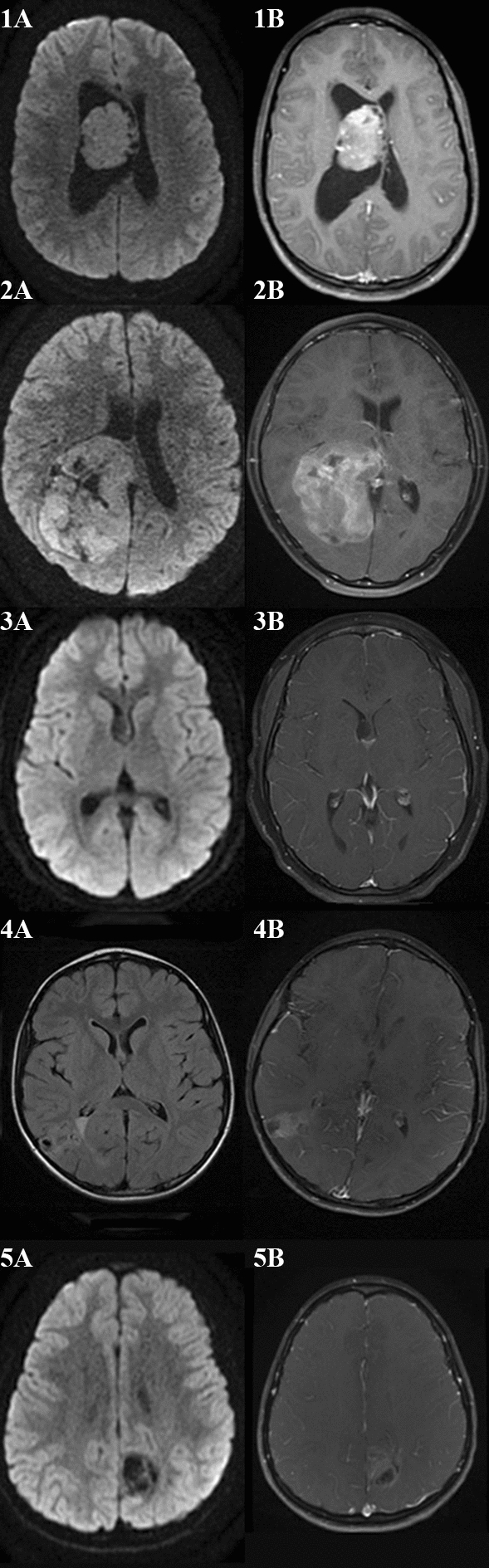
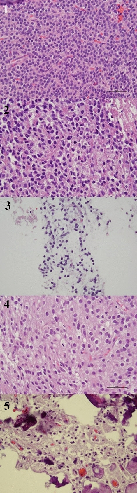
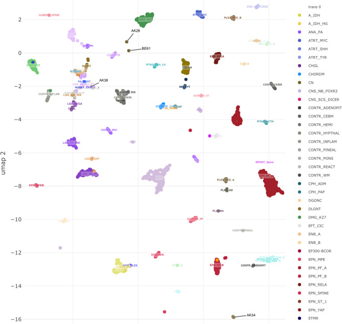
Results: Presenting symptoms included seizures (n = 2), syncope (n = 1), headache (n = 2), visual disturbances (n = 2) and emesis (n = 2). Tumor location included intraventricular (n = 2), intraventricular with parenchymal spread (n = 1), and extraventricular (n = 2). Magnetic resonance imaging demonstrated reduced diffusivity (2/5), signal abnormality on susceptibility-weighted sequences (3/5), and varying degrees of contrast enhancement (4/5). All patients underwent surgical resection alone. Recurrence occurred in four children that were treated with surgery (4/4), adjuvant radiation (2/4), and chemoradiation (1/4). Neuropathologic features included positivity for GFAP (4/5), synaptophysin (4/5), NSE (2/2), NeuN (4/4), and variable Ki-67 (< 1% to 15%). Next generation sequencing (3/5) and microarray (3/5) collectively were abnormal in four of five tumors. Methylation profiling was successfully performed on four of five samples which led to modification of diagnosis in two patients and the others were either unclassifiable or confirmatory with the histologic diagnosis. Mean time to follow up was 77 months (range 44-112 months). Mean progression free survival and overall survival were 24 months (range 6 to 52 months) and 100% respectively.

Conclusion: Neurocytomas are a rare clinical entity that warrants further investigation into molecular and pathologic prognosticating features. Methylation profiling may aid in differentiation of neurocytoma from other difficult to diagnose tumors who share similar histologic features.

Keywords: Atypical neurocytoma; Methylation; Neurocytoma; Pediatric brain tumor; Pediatric neurocytoma.


Conflict of interest statement

The authors declare no competing interests.

The authors have not disclosed any competing interests.

Figures

Fig. 1
Fig. 1
MRI of patient 1 demonstrates a central intraventricular tumor with reduced diffusivity (1A) and avid enhancement on post-gadolinium T1-weighted sequences (1B). MRI of patient 2 demonstrates a large posterior right lateral ventricle tumor with parietal and hemispheric parenchymal spread with reduced diffusivity (2A) and avid heterogenous contrast enhancement on post-gadolinium T1-weighted sequences (2B). MRI of patient 3 demonstrates a subtle intraventricular tumor adjacent to the septum pellucidum and right lateral ventricle on diffusion weighted sequences (3A) with minimal contrast enhancement likely reflecting choroid inferiorly on post-gadolinium T1-weighted sequences (3B). MRI of patient 4 demonstrates a right posterior temporoparietal mixed solid and cystic tumor on T2-weight fluid attenuated inversion recovery sequences (4A) with moderate enhancement on post-gadolinium T1-weighted sequences (4B). MRI of patient 5 demonstrates a mixed solid and cystic left posterior frontoparietal tumor without restricted diffusion on diffusion weighted sequences (5A) and moderate enhancement on post-gadolinium T1-weight sequences (5B).
Fig. 2
Fig. 2
Sample from patient 1 demonstrates highly cellular proliferation of uniform cells with round nuclei, stippled chromatin, moderately eosinophilic cytoplasm, and mitotic activity with 6 mitotic figures in 10 high-power fields. Sample from patient 2 demonstrates moderately cellular proliferation of polygonal cells with moderate nuclear pleomorphism, stippled chromatin, mitotic activity with 8 mitotic figures in 10 high-power fields, and necrosis. Sample from patient 3 demonstrates moderately cellular proliferation with round, regular nuclei, and stippled chromatin. Sample from patient 4 demonstrates moderately cellular proliferation with round nuclei showing perinuclear “halos” transitioning into more mature neuronal forms, extensive calcification without eosinophilic granular bodies or perivascular lymphocytic cuffing. Sample from patient 5 demonstrates moderately cellular proliferation with uniformly round nuclei, perinuclear “halo” formation, and extensive calcification.
Fig. 3
Fig. 3
Cluster analysis of the four patients with successful methylation testing are shown. Patient 1 (AK34) clustered with central neurocytoma (calibrated score=0.999). Patient 2 (AK28) did not cluster with any known entity. Patient 4 (AK38) clustered with ganglioglioma (calibrated score 0.927). Patient 5 clustered most closely to a ganglioglioma (calibrated score 0.766).

References

    1. Louis DN, Perry A, Wesseling P, Brat DJ, Cree IA, Figarella-Branger D, et al. The 2021 WHO classification of tumors of the central nervous system: a summary. Neuro Oncol. 2021;23:1231–1251. doi: 10.1093/neuonc/noab106. - DOI - PMC - PubMed
    1. Hassoun J, Gambarelli D, Grisoli F, Pellet W, Salamon G, Pellissier JF, et al. Central neurocytoma an electron-microscopic study of two cases. Acta Neuropathol. 1982;56:151–156. doi: 10.1007/BF00690587. - DOI - PubMed
    1. Hassoun J, Söylemezoglu F, Gambarelli D, Figarella-Branger D, von Ammon K, Kleihues P. Central neurocytoma: a synopsis of clinical and histological features. Brain Pathol. 1993;3:297–306. doi: 10.1111/j.1750-3639.1993.tb00756.x. - DOI - PubMed
    1. Tish S, Habboub G, Jones J, Ostrom QT, Kruchko C, Barnholtz-Sloan JS, et al. The epidemiology of central and extraventricular neurocytoma in the United States between 2006 and 2014. J Neurooncol. 2019;143:123–127. doi: 10.1007/s11060-019-03144-9. - DOI - PubMed
    1. Brant-Zawadzki G, Mathews MSG, Duma CM, Kim B, Chen P, Heinemann S, et al. Central neurocytoma: case report of an 81-year-old and histopathologic findings. Neuroradiol J. 2007;20:61–66. doi: 10.1177/197140090702000110. - DOI - PubMed