. 2022 Oct 1;79(10):1025-1035.
doi: 10.1001/jamaneurol.2022.2442.

Spatial Extent of Amyloid-β Levels and Associations With Tau-PET and Cognition

Collaborators, Affiliations

Spatial Extent of Amyloid-β Levels and Associations With Tau-PET and Cognition

Hazal Ozlen et al. JAMA Neurol. .

Abstract

Importance: Preventive trials of anti-amyloid agents might preferably recruit persons showing earliest biologically relevant β-amyloid (Aβ) binding on positron emission tomography (PET).

Objective: To investigate the timing at which Aβ-PET binding starts showing associations with other markers of Alzheimer disease.

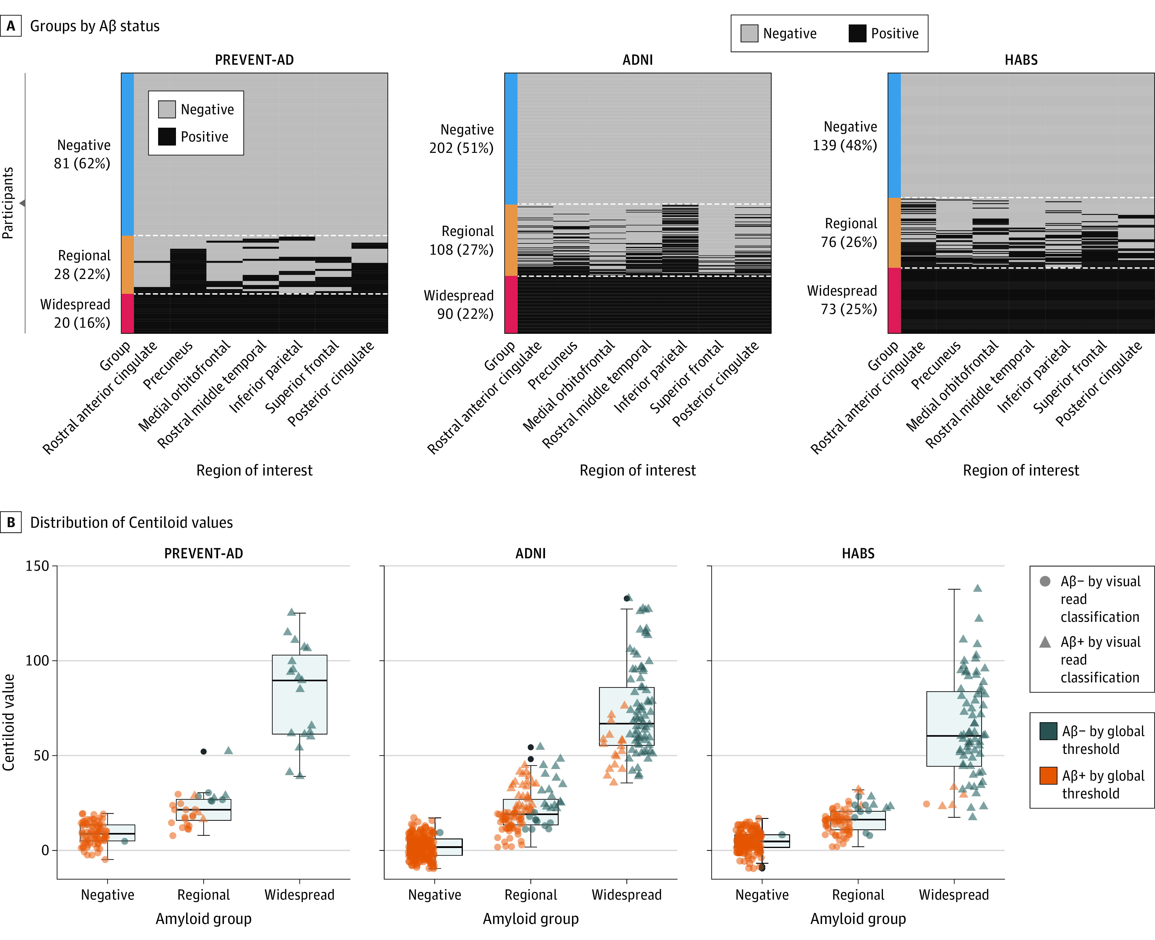
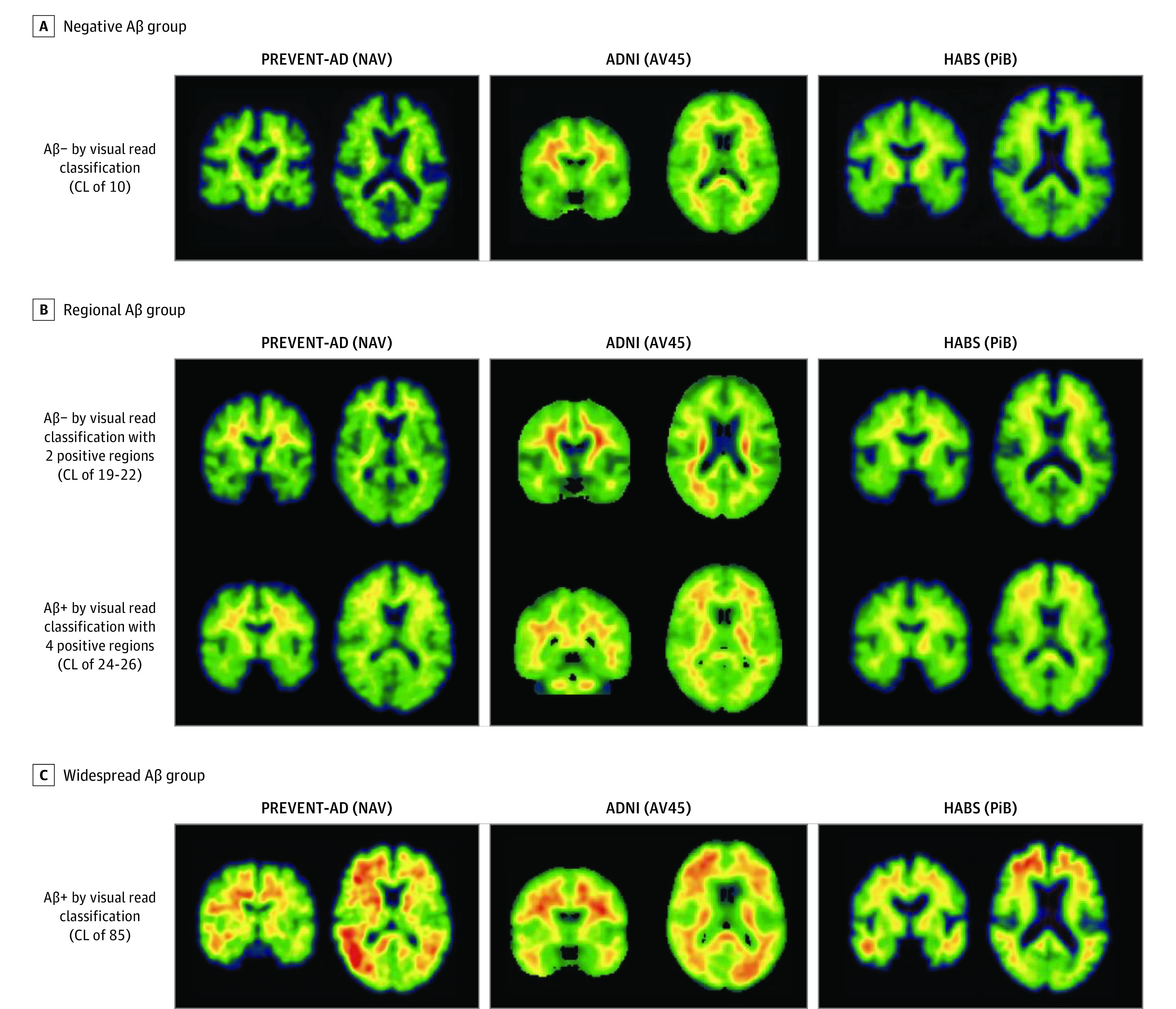
Design, setting, and participants: This longitudinal multicentric cohort study included 3 independent cohorts: Presymptomatic Evaluation of Experimental or Novel Treatments for Alzheimer Disease (PREVENT-AD) (data collected from 2012-2020), Alzheimer Disease Neuroimaging Initiative (ADNI) (data collected from 2005-2019), and Harvard Aging Brain Study (HABS) (data collected from 2011-2019). In a 3-tiered categorization of Aβ-PET binding spatial extent, individuals were assigned as having widespread Aβ deposition if they showed positive signal throughout a designated set of brain regions prone to early Aβ accumulation. Those with binding in some but not all were categorized as having regional deposition, while those who failed to show any criterion Aβ signal were considered Aβ-negative. All participants who were cognitively unimpaired at their first Aβ PET scan.

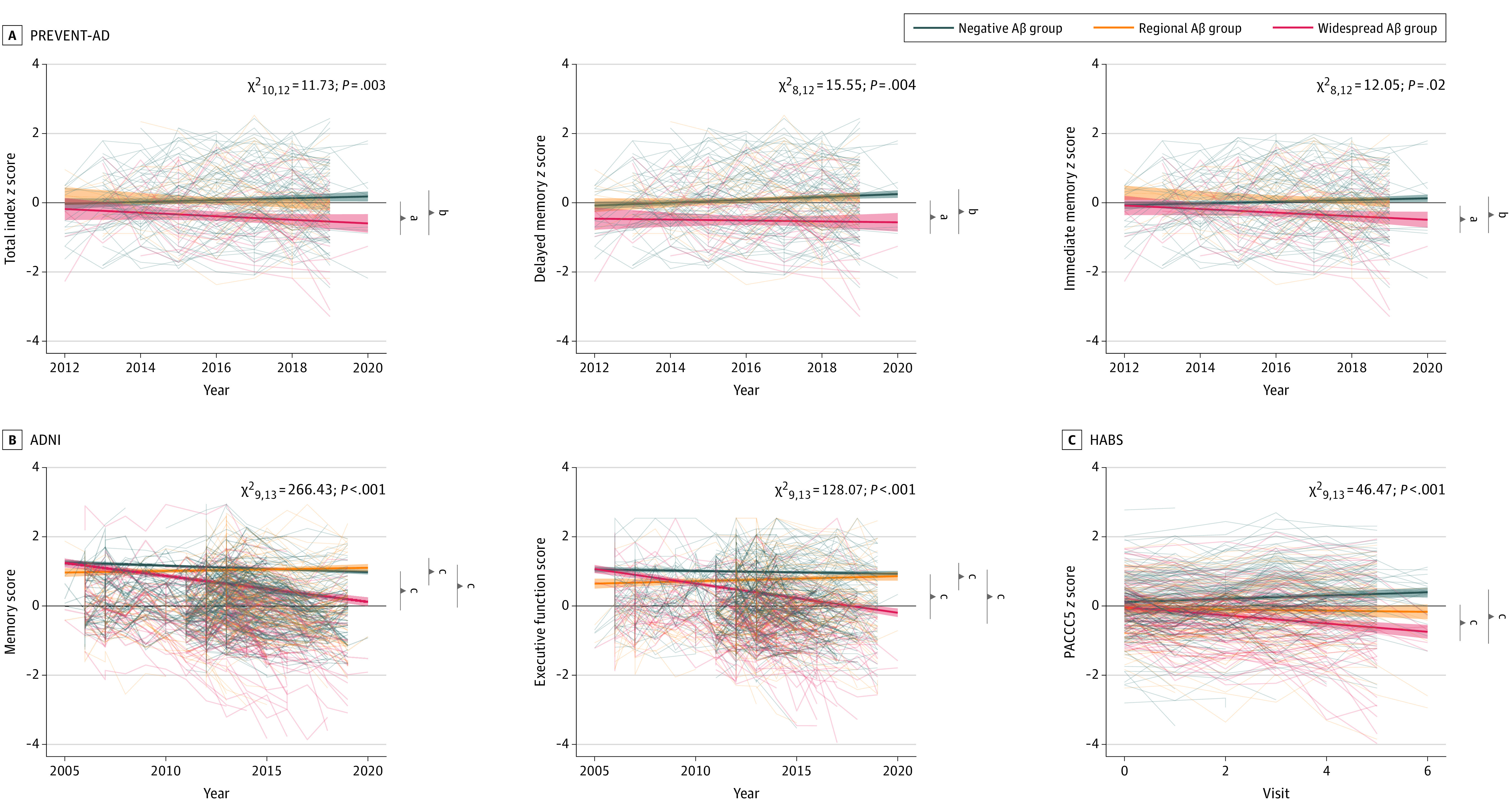
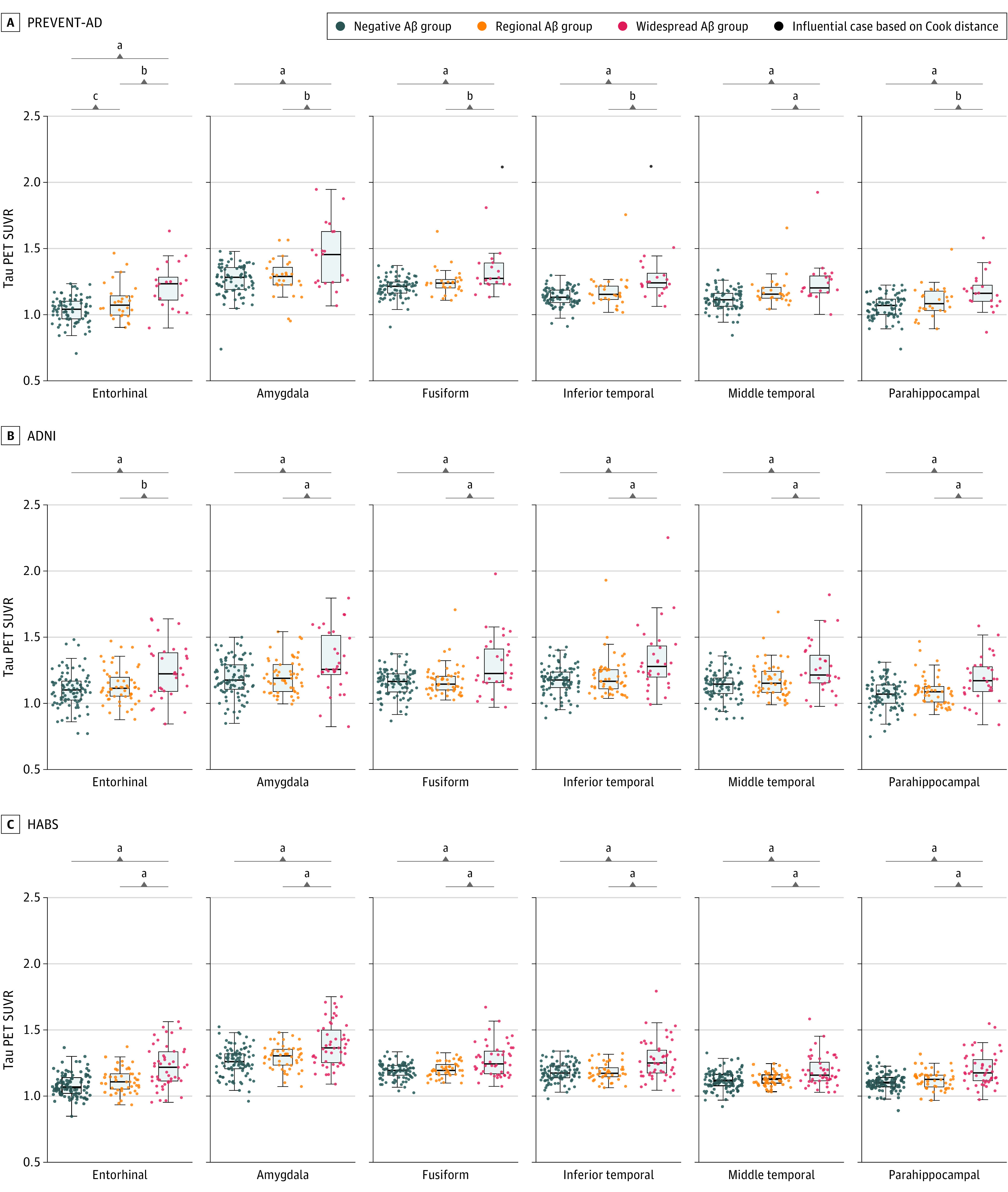
Main outcomes and measures: Differences in cerebrospinal fluid (CSF), genetics, tau-PET burden, and cognitive decline.

Results: A total of 817 participants were included, including 129 from the PREVENT-AD cohort (mean [SD] age, 63.5 [4.7] years; 33 [26%] male; 126 [98%] White), 400 from ADNI (mean [SD] age, 73.6 [5.8] years; 190 [47%] male; 10 [5%] Hispanic, 338 [91%] White), and 288 from HABS (mean [SD] age, 73.7 [6.2] years; 117 [40%] male; 234 [81%] White). Compared with Aβ-negative persons, those with regional Aβ binding showed proportionately more APOE ε4 carriers (18 [64%] vs 22 [27%] in PREVENT-AD and 34 [31%] vs 38 [19%] in ADNI), reduced CSF Aβ1-42 levels (F = 24 and 71), and greater longitudinal Aβ-PET accumulation (significant β = 0.019 to 0.056). Participants with widespread amyloid binding further exhibited notable cognitive decline (significant β = -0.014 to -0.08), greater CSF phosphorylated tau181 (F = 5 and 27), and tau-PET binding (all F > 7.55). Using each cohort's specified dichotomous threshold for Aβ positivity or a visual read classification, most participants (56% to 100%, depending on classification method and cohort) with regional Aβ would have been classified Aβ-negative.

Conclusions and relevance: Regional Aβ binding appears to be biologically relevant and participants at this stage remain relatively free from CSF phosphorylated tau181, tau-PET binding, and related cognitive decline, making them ideal targets for anti-amyloid agents. Most of these individuals would be classified as negative based on classical thresholds of Aβ positivity.


Conflict of interest statement

Conflict of Interest Disclosures: Ms Ozlen reported grants from Healthy Brains for Healthy Lives during the conduct of the study. Dr Soucy reported personal fees from Biogen Canada on behalf of company outside the submitted work. Dr Poirier reported grants from the Canadian Institutes of Health Research and J.L Levesque Foundation during the conduct of the study. No other disclosures were reported.

Figures

Figure 1.
Figure 1.. Defining the β-Amyloid (Aβ) Groups
A, Individuals were separated into 3 groups based on their Aβ status in 7 cortical regions: rostral anterior cingulate, precuneus, medial orbitofrontal, rostral middle frontal, inferior parietal, superior frontal, and posterior cingulate. According to the region-specific positivity, individuals who were Aβ-positive in all 7 regions were classified as the widespread Aβ deposition group; those who were positive in 1 to 6 regions were included in the regional Aβ group; while those who fell below the cutoff in all the regions were considered as Aβ-negative. B, The boxplots represent the distribution of the Centiloid values of the 3 Aβ groups across the Aβ-negative, regional, and widespread groups in all cohorts. Different shapes for the data points indicate the visual read classification, and the color categorizes participants as Aβ+ or Aβ− based on quantitative binary amyloid index using previously established global thresholds for each cohort. ADNI indicates Alzheimer Disease Neuroimaging Initiative; HABS, Harvard Aging Brain Study; PREVENT-AD, Presymptomatic Evaluation of Experimental or Novel Treatments for Alzheimer Disease.
Figure 2.
Figure 2.. Examples of β-Amyloid (Aβ) Uptake From Participants in the Negative, Regional, and Widespread Groups
Illustrative examples of Aβ standardized uptake value ratios positron emission tomography images from participants in the negative (A), regional (B), and widespread (C) Aβ groups in the 3 cohorts. All shown images in the negative group were negative based on visual read and had a Centiloid (CL) value of 10. We show examples of participants in the regional group who were positive on 2 regions and negative based on visual read and participants who were positive on 4 regions and positive based on visual read. In the Presymptomatic Evaluation of Experimental or Novel Treatments for Alzheimer Disease (PREVENT-AD) study cohort, the 2 and 4 regions positive were precuneus and posterior cingulate (2), plus rostral anterior cingulate and medial orbitofrontal (4). In the Alzheimer Disease Neuroimaging Initiative (ADNI) study cohort, the regions were rostral middle frontal and inferior parietal (2) and the 4 were the inferior parietal, precuneus, posterior cingulate, and medial orbitofrontal. In the Harvard Aging Brain Study (HABS) cohort, the regions were rostral anterior cingulate and medial orbitofrontal (2), plus rostral middle frontal and superior frontal (4). All images shown in the Widespread group were positive based on visual read and had a CL value of 85. The standardized uptake value ratios scales were restricted to 0 to 4 in PREVENT-AD, 0 to 3 in ADNI, and 0 to 3.5 in HABS. AV45 indicates 18F-AV-45; NAV, 18F-NAV-4694; PiB, 11C-Pittsburgh compound B.
Figure 3.
Figure 3.. Change in Cognition Over Time Between the 3 β-Amyloid (Aβ) Groups
Linear mixed-effect models were used to assess the main association of Aβ groups with longitudinal cognition, corrected for age, sex, and education. The analyses were anchored at the participants’ baseline visit date. Cognitive test scores for the Presymptomatic Evaluation of Experimental or Novel Treatments for Alzheimer Disease (PREVENT-AD) (A), Alzheimer Disease Neuroimaging Initiative (ADNI) (B), and Harvard Aging Brain Study (HABS) (C) cohorts were represented over time in the 3 different groups. The widespread Aβ group showed a greater decline in their cognition scores when compared with the 2 other groups in all cohorts. In both ADNI and HABS, the regional group showed a greater cognitive decline compared to the Aβ-negative group. PACC5 indicates the 5-item Preclinical Alzheimer’s Cognitive Composite. aP < .05. bP < .01. cP < .001.
Figure 4.
Figure 4.. Tau–Positron Emission Tomography (PET) Uptake Across the 3 β-Amyloid (Aβ) Groups
Six regions were chosen to represent areas of early tau-PET accumulation. Tau-PET scans were available for 129 Presymptomatic Evaluation of Experimental or Novel Treatments for Alzheimer Disease (PREVENT-AD) participants, 176 Alzheimer Disease Neuroimaging Initiative (ADNI) participants, and 195 Harvard Aging Brain Study (HABS) participants. A, In PREVENT-AD, the widespread Aβ group had elevated tau-PET signal when compared with Aβ-negative and regional Aβ groups across 6 regions. The regional Aβ group had elevated tau-PET binding only in the entorhinal cortex and middle temporal gyrus when compared with the Aβ-negative group. B and C, In both ADNI and HABS, the widespread Aβ group had elevated tau-PET signal compared with Aβ-negative and regional Aβ groups across all regions. Analyses were corrected for age and sex. SUVR indicates standardized uptake value ratios. aP < .001. bP < .01. cP < .05.

References

    1. Braak H, Braak E. Neuropathological stageing of Alzheimer-related changes. Acta Neuropathol. 1991;82(4):239-259. doi:10.1007/BF00308809 - DOI - PubMed
    1. Thal DR, Rüb U, Orantes M, Braak H. Phases of A beta-deposition in the human brain and its relevance for the development of AD. Neurology. 2002;58(12):1791-1800. doi:10.1212/WNL.58.12.1791 - DOI - PubMed
    1. Braak H, Del Tredici K. The pathological process underlying Alzheimer’s disease in individuals under thirty. Acta Neuropathol. 2011;121(2):171-181. doi:10.1007/s00401-010-0789-4 - DOI - PubMed
    1. Hardy J, Allsop D. Amyloid deposition as the central event in the aetiology of Alzheimer’s disease. Trends Pharmacol Sci. 1991;12(10):383-388. doi:10.1016/0165-6147(91)90609-V - DOI - PubMed
    1. Hardy J, Selkoe DJ. The amyloid hypothesis of Alzheimer's disease: progress and problems on the road to therapeutics. Science. 2002;297(5580):353-356. - PubMed