. 2022 Aug 30;119(35):e2209729119.
doi: 10.1073/pnas.2209729119. Epub 2022 Aug 22.

Malaria parasite evades mosquito immunity by glutaminyl cyclase-mediated posttranslational protein modification

Affiliations

Malaria parasite evades mosquito immunity by glutaminyl cyclase-mediated posttranslational protein modification

Surendra Kumar Kolli et al. Proc Natl Acad Sci U S A. .

Abstract

Glutaminyl cyclase (QC) modifies N-terminal glutamine or glutamic acid residues of target proteins into cyclic pyroglutamic acid (pGlu). Here, we report the biochemical and functional analysis of Plasmodium QC. We show that sporozoites of QC-null mutants of rodent and human malaria parasites are recognized by the mosquito immune system and melanized when they reach the hemocoel. Detailed analyses of rodent malaria QC-null mutants showed that sporozoite numbers in salivary glands are reduced in mosquitoes infected with QC-null or QC catalytically dead mutants. This phenotype can be rescued by genetic complementation or by disrupting mosquito melanization or phagocytosis by hemocytes. Mutation of a single QC-target glutamine of the major sporozoite surface protein (circumsporozoite protein; CSP) of the rodent parasite Plasmodium berghei also results in melanization of sporozoites. These findings indicate that QC-mediated posttranslational modification of surface proteins underlies evasion of killing of sporozoites by the mosquito immune system.

Keywords: glutaminyl cyclase; immune evasion; melanization; pyroglutamic acid; sporozoite.


Conflict of interest statement

The authors declare no competing interest.

Figures

Fig. 1.
Fig. 1.
Three-dimensional homology model, cyclase activity, and expression of P. falciparum QC. (A) Three-dimensional homology model of P. falciparum QC, generated against the resolved QC structures from C. papaya and Z. mobilis, and the 3D template structure of C. papaya QC. Structures are visualized using PyMOL. Helices, cyan; sheets, magenta; loops, brown. An overlay is shown of PfQC (green) and CpQC (red). (B) Visualization of the CpQC catalytic site (blue), composed of active-site residues F22, Q24, F67, E69, W83, W110, N155, W169, and K225 and the predicted PfQC (blue) catalytic site, based on the catalytic-site residues conserved with the CpQC catalytic site (F107, Q109, F152, E154, Y174, Y200, N269, F285, and K349; SI Appendix, Fig. S1D). (C) Cyclase activity of PfQC (relative fluorescence units, RFU), in an enzyme activity assay using recombinant wild-type (WT-PfQC; n = 4) and cyclase-dead PfQC (QCCD; n = 4), containing two point mutations in the active site, F107A and Q109A (***P < 0.0001). (D) QC expression in gametocytes, ookinetes, and sporozoites, visualized by staining with anti-cmyc antibodies of fixed Pbqc::cmyc parasites that express cmyc-tagged QC and mCherry. Nuclei are stained with Hoechst 33342. DIC, differential interference contrast. (Scale bars, 5 µm.)
Fig. 2.
Fig. 2.
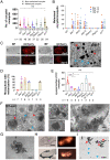
Melanization of oocysts and sporozoites in mosquitoes infected with QC-null mutants and reduced sg-sporozoite numbers. (A) Number of melanized and nonmelanized oocysts per mosquito (median + 95% CI; n, number of mosquitoes) infected with QC-null mutants (Δqc), QC-null mutants complemented with P. berghei or P. falciparum QC [Pbqc(c) and Pfqc(c)], or infected with a QC catalytically dead mutant (qcCD). At least three replicates [except Pbqc(c): one experiment and qcCD in duplicate] were pooled for statistical analysis. ns, not significant [Mann–Whitney U test; significance relative to WT; P values: Δqc1 0.159, Δqc2 0.174, ΔqcG 0.188, Pbqc(c) 0.360, Pfqc(c) 0.079, and qcCD 0.062]. (B) Number of melanized oocysts per mosquito (day 14, 17, and 21) (n = 15 to 20) infected with qc mutants. Number of mosquitoes on day 14, 17, and 21: WT (18, 21, and 18); Δqc1 (10, 32, and 5); Δqc2 (16, 10, and 12); Δqc-G (14, 18, and 5); Pbqc(c) (28, 11, and 18); Pfqc(c) (16, 19, and 22); qcCD (17, 24, and 15). At least three replicates [except Pbqc(c): one experiment and qcCD in duplicate] were pooled for statistical analysis and error bars represent the median with 95% CI. (C) Melanized oocyst in mosquito midguts (day 14) infected with QC-null mutants (Δqc). Bright-field (BF) and fluorescence images of mCherry-expressing oocysts. (Scale bars, 200 µm.) (C, Right) Melanized (red arrowheads) and nonmelanized (blue arrowheads) oocysts in a Δqc1-infected mosquito (day 14). Nonmelanized oocysts are shown in blue circles. (Scale bar, 100 µm.) (D) Percentage of mosquitoes with melanized oocysts (n, number of experiments; 30 to 40 mosquitoes per experiment; day 14) infected with different qc mutant parasites (see A for mutant names). At least three replicates [except Pbqc(c): one experiment] were pooled for statistical analysis. Data are represented as mean ± SD. (E) Number of sg-sporozoites per mosquito (n, number of experiments; 60 to 80 mosquitoes per experiment) infected with qc mutants. Mean and SD [Δqc: three to six experiments; WT: six experiments; Pbqc(c): one experiment; other mutants: two to four experiments]. **P < 0.005, *P < 0.05; ns, not significant [Mann–Whitney U test; significance relative to WT; P values: Δqc1 0.0095, Δqc2 0.004, ΔqcG 0.024, Pfqc(c) 0.262, qc::cmyc 0.191, and qcCD 0.071]. (F) Examples of QC-null oocysts showing melanization of sporozoites inside or in the process of egress (Left and Middle); red arrows: melanized oocysts; blue arrows: WT-like oocysts. (F, Inset) WT oocyst with sporulation (day 14). (Scale bars, 20 µm.) (F, Right) Melanized QC-null hemocoel sporozoites (abdominal region; day 21). (Scale bar, 25 µm.) (G) Cluster of melanized (dark-colored, enlarged) sporozoites in the hemocoel of a QC-null–infected mosquito (day 21). (Scale bar, 10 µm.) (H) Bright-field and fluorescence images of salivary glands (day 21) from mosquitoes infected with WT (Upper) and QC-null parasites (Lower), expressing mCherry (Right). Melanized, mCherry-negative sporozoites cluster at the periphery of salivary glands. (Scale bars, 50 µm.) (I) Partially crushed salivary gland (day 21) of a QC-null–infected mosquito showing melanized (red arrowheads) and nonmelanized (blue arrowheads) sporozoites. (Scale bar, 10 µm.)
Fig. 3.
Fig. 3.
SEM-EDX analysis of melanized and nonmelanized QC-null oocysts and sporozoites. (A) SEM (Left) and bright-field (Right) identification and selection of oocysts (5174) and sporozoites (5173) in a midgut of an A. stephensi mosquito infected with PbΔqc1 (day 14). (B) EDX line scan of the elements carbon (blue) and sulfur (orange) together with SEM images (counts per second, CPS) showing the presence of sulfur in melanized oocysts (Left; dark-colored oocyst in 5174; A) and in melanized, dark-colored sporozoites (Right; 5173; A). Black lines represent EDX line scan positions. Yellow arrowheads indicate melanized black oocysts, and white arrowheads indicate nonmelanized oocysts (5174). Cluster of melanized sporozoites (5173). (C) EDX mapping analysis of carbon and sulfur together with an SEM image of melanized sporozoites (5173; A).
Fig. 4.
Fig. 4.
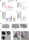
Melanization of oocysts and sporozoites of P. falciparum QC-null mutants. (A) Number of melanized and nonmelanized oocysts per mosquito in A. stephensi mosquitoes (n, number of mosquitoes) infected with QC-null mutants (Δqc) or WT parasites. Error bars represent the median ± 95% CI (Δqc: two experiments; WT: four experiments). ns, not significant (Mann–Whitney U test; statistical significance is shown relative to WT; P values: Δqc1 0.333 and Δqc2 0.531). (B) Number of sg-sporozoites per mosquito (n, number of experiments; 60 to 80 mosquitoes per experiment), infected with QC-null mutants (Δqc) or WT parasites. Mean and SD [two experiments for QC-null mutants (Δqc) and four experiments for WT]. ns, not significant (Mann–Whitney U test; statistical significance is shown relative to WT; P values: Δqc1 0.133 and Δqc2 0.133). (C) Number of melanized oocysts per individual mosquito on days 11, 15, and 21 after infection with QC-null mutants or WT parasites (n, number of mosquitoes). The horizontal bars indicate medians (two to four experiments). (D) Percentage of mosquitoes with melanized oocysts (n, number of experiments; 30 to 40 mosquitoes per experiment) at day 21 after infection with QC-null mutants or WT parasites. Data are represented as mean ± SD. (E) Melanized (dark-colored) oocysts in midguts of mosquitoes (day 11) infected with QC-null mutants. No melanized oocysts were observed in WT parasites. (Scale bars, 200 µm.) (F) Examples of (partly) melanized QC-null oocysts (M, day 15) showing melanization of sporozoites still inside or in the process of oocyst egress and an oocyst with typical features of WT sporozoite formation (WT-like). (Scale bars, 20 µm.) (G) Melanized (red circles) and nonmelanized, WT-like (purple circles) QC-null sporozoites obtained from a salivary gland (under a coverslip) isolated from an infected mosquito (day 21). (Scale bars, 10 µm.)
Fig. 5.
Fig. 5.
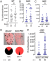
Silencing of mosquito immune responses results in increased sporozoite numbers in salivary glands of mosquitoes infected with P. berghei QC-null mutants. (A) Sporozoite density (luciferase activity) in individual mosquito thoraces at day 23 p.i. after systemic injection of polystyrene beads into the thorax (day 12 p.i.) of mosquitoes infected with WT parasites (n, number of mosquitoes). ns, not significant (Mann–Whitney U test, P > 0.05). The horizontal bars indicate medians. (B) Sporozoite density (luciferase activity in individual mosquito thoraces; day 23 p.i.) after systemic injection of polystyrene beads into the thorax (day 12 p.i.) of QC-null (Δqc2)–infected mosquitoes (n, number of mosquitoes). **P < 0.01 (Mann–Whitney U test). The horizontal bars indicate medians. (C) Sporozoite density (luciferase activity in individual mosquito thoraces; day 23 p.i.) after injection of dsRNA for LRIM1 (ASTE000814) (dsLRIM1) or LacZ (dsLacZ; control) (day 12 p.i.) of QC-null mutant (Δqc2)–infected mosquitoes (n, number of mosquitoes). ns, not significant (Mann–Whitney U test, P > 0.05). The horizontal bars indicate medians. (D) Representative midguts and percentage of mosquitoes with melanized (black) and nonmelanized (red) parasites (23 d p.i.) after injection of dsRNA for CLIPA8 (ASTE009395) (dsCLIPA8) or LacZ (dsLacZ; control) (day 12 p.i.) of QC-null mutant (Δqc2)–infected mosquitoes (n, number of mosquitoes). ****P < 0.0001 (χ2 test). (E) Sporozoite density (luciferase activity in individual mosquito thoraces; day 23 p.i.) after injection of dsCLIPA8 or dsLacZ (control) (day 12 p.i.) of QC-null mutant (Δqc2)–infected mosquitoes (n, number of mosquitoes). ****P < 0.0001 (Mann–Whitney U test). The horizontal bars indicate medians.
Fig. 6.
Fig. 6.
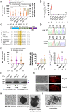
Absence of melanization of QC-null oocysts in the absence of sporozoite egress, and melanization of oocysts and sporozoites that express CSP with a mutated QC-target glutamine. (A) Number of melanized and nonmelanized oocysts per mosquito (median ± 95% CI; n = 40 to 50) infected with a QC-null mutant [Δqc1(-sm)], WT and QC-null mutants ΔqcΔcsp, ΔqcΔrom3 (no sporozoite formation), ΔqcΔecp1, ΔqcΔcrmp4 (no sporozoite egress), and ΔqcΔtrap (sporozoite egress). n, number of mosquitoes. y axis (Right): percentage of mosquitoes with melanized oocysts [three experiments for all mutants except for Δqc1(-sm), n = 4]. ns, not significant [Mann–Whitney U test; significance relative to WT; P values: ΔqcΔcsp 0.003, ΔqcΔrom3 0.968, ΔqcΔecp1 0.184, ΔqcΔcrmp4 0.216, ΔqcΔtrap 0.895, and Δqc(-sm) 0.075]. (B) Number of sg-sporozoites per mosquito infected with mutants as shown (Left) (n, number of experiments; 60 to 80 mosquitoes per experiment). Data are represented as mean ± SD. ns, not significant [Mann–Whitney U test; significance relative to WT; P value: Δqc(-sm) 0.071]. (C) Schematic showing different regions of CSP. Yellow: region 1 (RI). Dark yellow box: the conserved glutamine in R1 (KLKQP). R1 is highly conserved and contains the cleavage site (41). CTD, C-terminal domain; NTD, N-terminal domain; SP, signal peptide. (D) Sanger-sequence DNA chromatograms of the PCR fragment of P. berghei CSP RI amplified from WT and cspmut genomic DNA, confirming the replacement of glutamine with alanine (Q92A; highlighted) in cspmut. (E, Left) Number of melanized and nonmelanized oocysts per mosquito in A. stephensi mosquitoes (n, number of mosquitoes) infected with CSP Q92A mutants (CSPmut1 and CSPmut2) or WT parasites. Error bars represent the median ± 95% CI (four to six experiments). ns, not significant (Mann–Whitney U test; statistical significance is shown relative to WT; P values: cspmut1 0.439 and cspmut2 0.264). (E, Middle) Percentage of mosquitoes with melanized oocysts (n, number of experiments; 30 to 40 mosquitoes per experiment; day 16) infected with cspmut. No melanized oocysts in WT-infected mosquitoes. Data are represented as mean ± SD. (E, Right) Number of sg-sporozoites per mosquito (day 21; n, number of experiments; 60 to 80 mosquitoes per experiment) infected with WT, cspmut1, and cspmut2. Data are represented as mean ± SD (n, number of experiments). ns, not significant (Mann–Whitney U test; significance relative to WT; P values: cspmut1 0.073 and cspmut2 0.073). (F) Western analysis of sporozoite lysates showing CSP expression/processing in WT and cspmut, expressing mutated CSP (Q92A), using antibody 3D11, recognizing CSP repeats. Processing results in a full-length (55 kDa) and a processed form (45 kDa) (39, 48). WT and cspmut sporozoites incubated with or without heparin at 37 °C or room temperature (RT; 10 min) as CSP also undergoes processing when in contact with heparan sulfate proteoglycans (21). (G) Midguts of mosquitoes with mCherry-expressing cspmut oocysts, showing melanized (dark-colored) oocysts. (Scale bars, 200 µm.) (H) Bright-field light-microscope images of cspmut oocysts (day 21) showing WT-like (WT-ooc) and melanized oocysts. (H, Right) Melanized sporozoites during egress. (Scale bars, 20 µm.) (I) Melanized cspmut sporozoites in the hemocoel (abdominal region; day 21). (Scale bar, 10 µm.)
Fig. 7.
Fig. 7.
Proposed model of immune evasion of WT oocysts and sporozoites by posttranslational modification of proteins by QC. A mechanism of immune evasion by Plasmodium in the mosquito host is proposed in which QC modification of Plasmodium-target proteins prevents melanization of sporozoites (spz) as they come in contact with mosquito hemolymph. QCs posttranslationally modify proteins with N-terminal glutamine or glutamic acid to a cyclic pyroglutamic acid. Sporozoites and rupturing oocysts lacking QC activity (Δqc, qcCD) or expressing CSP with a mutated glutamine (in R1, cspmut) are recognized and melanized (indicated as black oocyst and sporozoites). Sporozoites of Δqc, qcCD, and cspmut are also melanized in the hemocoel. Sporozoites that escape melanization and successfully invade the salivary gland are protected from mosquito immune attack. Sporozoite melanization is a major mechanism eliminating Δqc sporozoites, because disrupting melanization by silencing CLIPA8 significantly increases the number of viable Δqc sporozoites. This indicates that sporozoite melanization is not activated in response to parasites that are already dead or irreversibly damaged. Mutating a single QC-target glutamine of CSP, the major sporozoite surface protein, also results in melanization of sporozoites. pGlu formation at this glutamine of WT sporozoites requires processing of CSP. CSP cleavage in R1 occurs at the sporozoite surface. We propose a working model in which proteolytic cleavage of CSP in R1 (KLKQP), between lysine and glutamine, generates an N-terminal glutamine that is posttranslationally modified to pGlu by the enzymatic activity of QC, and this posttranslational modification increases the probability that sporozoites will reach the salivary gland unharmed. CT, C-terminal fragment; NT, N-terminal fragment; RR, repeat region; SP, signal peptide (amino acids in R1 are shown as single-letter codes).

References

    1. Wintjens R., et al. , Crystal structure of papaya glutaminyl cyclase, an archetype for plant and bacterial glutaminyl cyclases. J. Mol. Biol. 357, 457–470 (2006). - PubMed
    1. Booth R. E., Lovell S. C., Misquitta S. A., Bateman R. C. Jr., Human glutaminyl cyclase and bacterial zinc aminopeptidase share a common fold and active site. BMC Biol. 2, 2 (2004). - PMC - PubMed
    1. Schilling S., Wasternack C., Demuth H. U., Glutaminyl cyclases from animals and plants: A case of functionally convergent protein evolution. Biol. Chem. 389, 983–991 (2008). - PubMed
    1. Vijayan D. K., Zhang K. Y. J., Human glutaminyl cyclase: Structure, function, inhibitors and involvement in Alzheimer’s disease. Pharmacol. Res. 147, 104342 (2019). - PubMed
    1. Saido T. C., et al. , Dominant and differential deposition of distinct beta-amyloid peptide species, A beta N3(pE), in senile plaques. Neuron 14, 457–466 (1995). - PubMed

Publication types