Review
. 2022 Aug;30(3):383-407.
doi: 10.1016/j.mric.2022.04.004.

Magnetic Resonance Imaging for Spine Emergencies

Affiliations
Review

Magnetic Resonance Imaging for Spine Emergencies

Jeannette Mathieu et al. Magn Reson Imaging Clin N Am. 2022 Aug.

Abstract

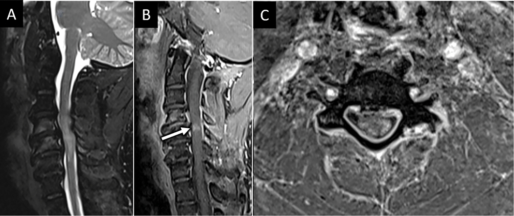
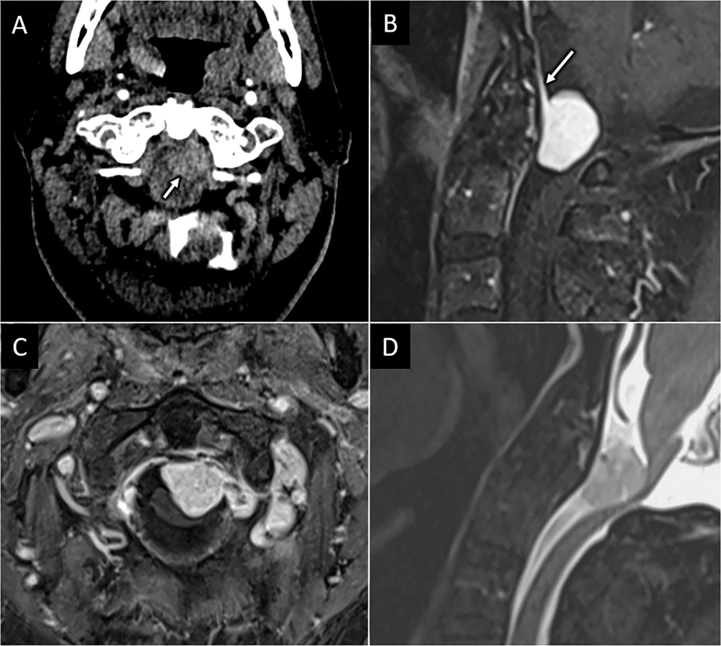
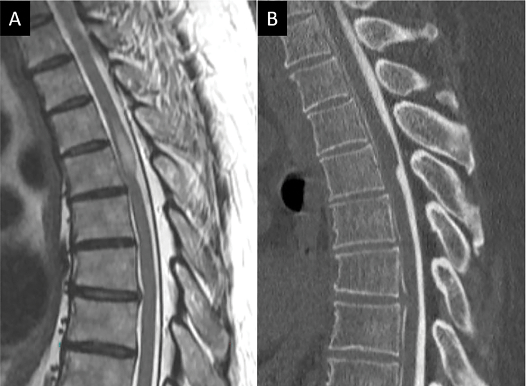
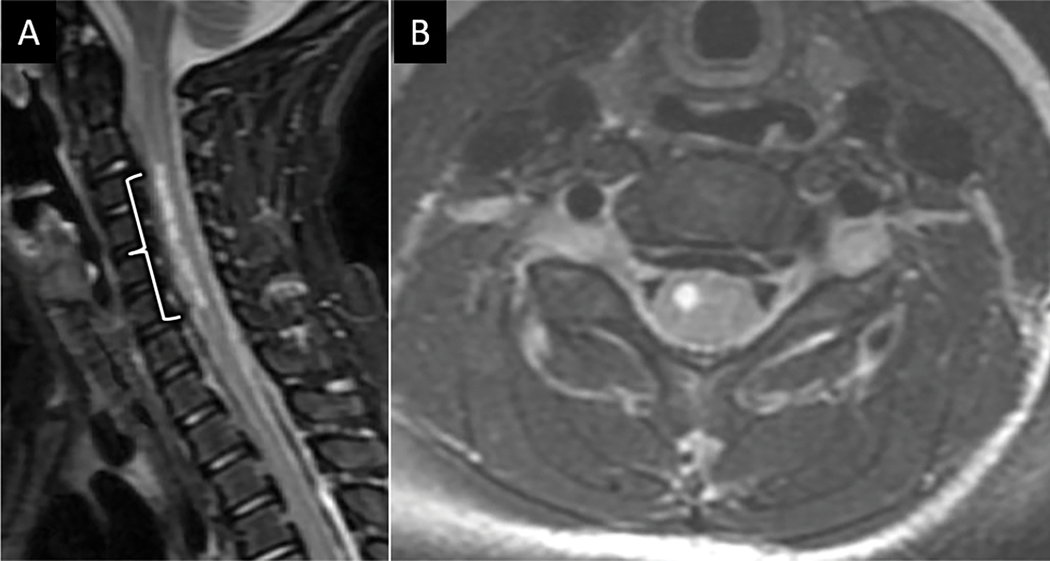
This article is devoted to the MR imaging evaluation of spine emergencies, defined as spinal pathologic conditions that pose an immediate risk of significant morbidity or mortality to the patient if not diagnosed and treated in a timely manner. MR imaging plays a central role in the timely diagnosis of spine emergencies. A summary of MR imaging indications and MR imaging protocols tailored for a variety of spinal emergencies will be presented followed by a review of key imaging findings for the most-encountered emergent spine pathologic conditions. Pathologic conditions will be broadly grouped into traumatic and atraumatic pathologic conditions. For traumatic injuries, a practical and algorithmic diagnostic approach based on the AO Spine injury classification system will be presented focused on subaxial spine trauma. Atraumatic spinal emergencies will be dichotomized into compressive and noncompressive subtypes. The location of external compressive disease with respect to the thecal sac is fundamental to establishing a differential diagnosis for compressive emergencies, whereas specific patterns of spinal cord involvement on MR imaging will guide the discussion of inflammatory and noninflammatory causes of noncompressive myelopathy.

Keywords: MR imaging; Myelitis; Myelopathy; Spinal cord; Spinal cord injury.


Conflict of interest statement

Disclosure The authors have nothing to disclose

Figures

FIGURE 1:
FIGURE 1:
Hyperextension injury with unstable anterior tension band (ATB) disruption. 85-year-old man presents with left sided weakness after ground level fall and headstrike. Sagittal CT image (arrow in A) shows traumatic anterolisthesis at C6-C7 with small anteroinferior avulsion corner fracture of C6 (arrow). Sagittal FS T2W (B) and sagittal T2 SPACE (C) MR images confirm complete disruption of the C6-C7 anterior discoligamentous complex. MRI also reveals long segment severe spinal canal stenosis and cord compression with dorsal epidural hematoma (arrows in C). Focal ligamentum flavum tear at C5–6 is best seen on sagittal T2 SPACE image (arrowhead in C).
FIGURE 2:
FIGURE 2:
Hyperextension injury with anterior longitudinal ligament (ALL) disruption. 70-year-old female presents with upper extremity paresthesia following low speed pedestrian versus automobile injury. A) Sagittal CT image of the cervical spine shows a small and minimally displaced anteroinferior avulsion corner fracture at C3 (dotted square with zoom in view in the bottom right inset in A). B) Sagittal T2 FS MR imaging of the upper cervical spine in the same patient shows prominent prevertebral fluid collection with focal disruption of the ALL (arrow in B) with otherwise intact anterior discoligamentous complex.
FIGURE 3:
FIGURE 3:
MRI is sensitive for mild vertebral compression injuries. Sagittal CT image (A) in a patient who presented with neck pain after a fall reveals mild superior endplate compression fractures at C6 and C7 (arrows in A), which are easily seen on sagittal T2W MRI (B) where linear subchondral marrow edema underlying the superior endplates conspicuously identifies the acute fractures.
Figure 4:
Figure 4:
MRI reveals unsuspected ligamentous posterior tension band disruption. Sagittal CT image (A) for a 48-year-old patient suffering a fall reveals mild anterior compression and endplate fractures at T5 and T6 vertebral levels. Sagittal T2W MR image (B) at the same levels reveals complete disruption of the supraspinous ligament, interspinous ligament, and ligamentum flavum at T5-T6 (arrows in B).
Figure 5:
Figure 5:
CT and MRI depictions of Chance fracture. Sagittal CT (A) depicts linear fracture through the spinous process in a 68-year-old female with seatbelt injury and low back pain. B) 3D reconstruction of the CT images clearly depicts transverse fracture through the entire L1 posterior elements (arrows in B) consistent with an AOSpine type B1 injury. Sagittal T2W MR image in the same patient shows marrow edema and transverse distracted fracture line corresponding to osseous posterior tension band fracture (white dotted line in C).
Figure 6:
Figure 6:
MRI confirms suspected posterior ligamentous complex injury. Sagittal FS T2W (A) and T2 SPACE (B) MR images obtained for a 33-year-old restrained passenger following motor vehicle collision reveals abnormal edema along the posterior ligamentous complex at C5-C6 (arrow in A and B).
Figure 7:
Figure 7:
Sagittal and axial MRI-based grading schema for assessment of acute traumatic spinal cord injury.
Figure 8:
Figure 8:
Lumbar septic facet arthritis with spinal epidural abscess. Axial T1W postcontrast (A) and T2W (B) MR images from a 54-year-old male with diabetes mellitus type 2 reveals right L5-S1 peri-facet joint soft tissue enhancement and multiloculated abscess (dotted circle) consistent with septic facet arthritis. There is associated peripherally enhancing and T2 hyperintense fluid collection in the dorsal epidural space of the lower lumbar spine (arrow in A and B). Sagittal T1W postcontrast MR image shows longitudinal extension of this collection to upper lumbar levels (arrows in C). Sagittal diffusion weighted (D) and apparent diffusion coefficient (E) images confirm abnormal reduced diffusion corresponding to the fluid collection consistent with purulent abscess (arrows in D and E).
Figure 9:
Figure 9:
DOM with epidural phlegmon resulting in spinal canal narrowing and spinal cord compression. Sagittal T1W-post contrast (A) and T2W (B) MR images centered at the mid-cervical spine show characteristic features of C4–5 and C5–6 DOM with prominent, solidly enhancing ventral epidural phlegmon (arrow in A and B).
Figure 10:
Figure 10:
Tuberculous spondylitis. Sagittal T2W FS (A) and T1W-post contrast MR images from a 31-year-old male with insidious cough, fever and weight loss reveals abnormal edema and enhancement centered at the T5 spina level notable for anterior subligamentous phlegmon extension (arrows in A and B) and relative preservation of adjacent intervertebral discs. Subsequent percutaneous biopsy confirmed mycobacterial infection.
Figure 11:
Figure 11:
Pathologic versus osteoporotic compression fractures. Sagittal T2W (A), T1W (B), T1W-post contrast (C), DWI (D), and ADC map (E) of the lumbar spine from a 59-year-old female with low back pain and breast cancer show L4 compression deformity with masslike dorsal bowing of the posterior vertebral wall encroaching on the spinal canal. Mass-like extra-osseous enhancement extending from the posterior L4 vertebral body has abnormal reduced diffusion (arrow in D and E) consistent with hypercellular tumor. Similar series of MR images (F-J) in a separate 82-year-old osteoporotic female shows L4 vertebral compression fracture with non-masslike enhancement, posterior bony retropulsion, and facilitated rather than reduced diffusion within the L4 vertebral body, consistent with osteoporotic fracture.
FIGURE 12:
FIGURE 12:
MRI features of spontaneous epidural hematoma. Axial CT (A) and T2W MR (B) images at the mid-thoracic level from a 41-year-old man presenting with acute onset loss of sensory and motor function to his bilateral lower extremities immediately following bowel movement reveal intrinsic hyperdense and T2 heterogenous expansion of the right dorsolateral epidural space with resultant severe spinal canal stenosis and spinal cord compression (arrow in A and B). C) Sagittal T2W image from the same patient clearly depicts the dorsal epidural location of this collection with uplifting of the dural membrane (arrowheads in C). Internal T2 hypointensity within the hematoma corresponds to paramagnetic T2 shortening from blood products.
Figure 13:
Figure 13:
Spondylotic myelopathy. A) Sag T2W FS MR image from a 46-year-old man with lower extremity weakness and fall shows severe degenerative spinal canal stenosis at the C4-C5 level with abnormal intramedullary spinal cord T2 hyperintensity centered at this level. Sagittal (B) and axial (C) T1W-post contrast images reveal transverse pattern of “pancake” like enhancement (arrow in B) which primarily involves peripheral white matter columns, consistent with spondylotic myelopathy.
Figure 14:
Figure 14:
Intradural compressive meningioma. Axial CT angiogram (A) performed for a 78-year-old male presenting with upper extremity paresthesia and weakness after fall shows subtle enhancing mass within the spinal canal at the C1 level (arrow in A). Sagittal (B) and axial (C) T1W FS-post contrast MR images show an intradural solidly enhancing mass with broad dural attachment and dural tail (arrow in B). Sagittal T2W FS MR image (D) shows severe spinal canal stenosis at C1 with cervicomedullary junction compression.
Figure 15:
Figure 15:
Arachnoid web with compressive myelopathy. 54-year-old man presents with severe upper thoracic pain. A) Sagittal T2W MR image centered at the upper thoracic spine reveals concave indentation along the dorsal spinal cord resembling the profile of a surgical scalpel (i.e. “scalpel” sign) with intramedullary spinal cord T2 hyperintense edema above the level of spinal cord compression. Subsequent CT-myelogram (B) excluded spinal cord herniation and confirmed diagnosis of spinal arachnoid web.
Figure 16:
Figure 16:
Enterovirus related acute flaccid myelitis. 3-year-old boy with ten-day loss of movement in the right arm in setting of upper respiratory illness. Sagittal (A) and axial (B) T2W MR images show abnormal T2-hyperintensity spanning multiple cervical vertebral segments centered in the right frontal horn of the spinal cord.
Figure 17:
Figure 17:
Neuromyelitis optica spectrum disorder (NMOSD) and myelin oligodendrocyte glycoprotein antibody disease (MOGAD) related myelopathies. Sagittal (A) and axial (B) T2W MR images from a 51-year-old women presenting with acute onset lower extremity weakness and numbness reveals longitudinally extensive centromedullary pattern of spinal cord edema commonly seen with NMO. Sagittal (C) and axial (D) T2W MR images from a different 15-year-old patient with recurrent hospitalizations for sensory and motor deficits reveals patchy T2 edema in the cervical spine with central gray-matter involvement resembling the letter “H” (D).
Figure 18:
Figure 18:
Spinal neurosarcoidosis. Sagittal T2W (A) MR image of the cervical spine from a 51-year-old man with progressive numbness, spasticity, and pain reveals multifocal patchy peripheral spinal cord T2 hyperintensity. Sagittal (B) and axial (C) T1W-post contrast MR images demonstrate associated “trident” pattern of dorsal pial and infiltrative subpial spinal cord enhancement (arrows in C), a pattern typical of spinal neurosarcoidosis.
Figure 19:
Figure 19:
Acute anterior spinal cord infarct. Sagittal (A) and axial (B) T2W FS MR images from a 68-year-old man with acute onset upper extremity weakness following aortic aneurysm repair shows ventral frontal-horn predominant T2 hyperintensity. Sagittal DWI (C) and ADC map (D) from the same exam shows abnormal reduced diffusion (arrow in C and D) corresponding with T2 signal abnormality consistent with acute anterior spinal artery infarct.
Figure 20:
Figure 20:
Spinal dural arteriovenous fistula (sdAVF). Sagittal (A) and axial (B) T2W MR images from a 52-year-old female with rapidly progressive bilateral lower extremity weakness and urinary incontinence reveals abnormal centromedullary pattern of T2 hyperintensity in the lower spinal cord and conus. Axial T1W-post contrast image of the cauda equina (C) shows abnormal nerve root hyperenhancement. Sagittal T2W image of the thoracic spine (D) shows abnormal dilated vascular flow voids along the dorsal surface of the spinal cord (arrowheads in D). 3-dimensional reconstruction from digital subtraction angiography with selective injection of the right T9 intercostal artery reveals multiple small branches coalescing at a dilated vein within the T10-T11 neuroforamen (arrow in E) with dilated venous outflow extending cranial and caudal along the dorsum of the spinal cord, consistent with sdAVF.
Figure 21:
Figure 21:
Dorsal column tractopathy related to Vitamin B12 deficiency. Sagittal (A) and axial (B) T2W MR images of the cervical spine from a 36-year-old vegetarian female with low vitamin B12 levels and 1 week history of bilateral hand and lower extremity numbness and gait instability demonstrates longitudinally extensive dorsal column T2 hyperintensity (arrows in A). Axial post-contrast T1W MRI (C) shows no associated abnormal enhancement.
Figure 22:
Figure 22:
Guillain Barre Syndrome. Sagittal (A) and axial (B) T1W-post contrast images of the lumbar spine show diffuse thickening and hyperenhancement of the ventral motor nerve rootlets in this 42-year-old female presenting with 1 month of rapidly progressive ascending weakness following a gastrointestinal illness.

References

    1. Daffner RH, Hackney DB. ACR Appropriateness Criteria on suspected spine trauma. J Am Coll Radiol. Nov 2007;4(11):762–75. doi:10.1016/j.jacr.2007.08.006 - DOI - PubMed
    1. Shah LM, Ross JS. Imaging of Spine Trauma. Neurosurgery. Nov 2016;79(5):626–642. doi:10.1227/NEU.0000000000001336 - DOI - PubMed
    1. Talbott JF, Huie JR, Ferguson AR, Bresnahan JC, Beattie MS, Dhall SS. MR Imaging for Assessing Injury Severity and Prognosis in Acute Traumatic Spinal Cord Injury. Radiol Clin North Am. Mar 2019;57(2):319–339. doi:10.1016/j.rcl.2018.09.004 - DOI - PubMed
    1. Arce D, Sass P, Abul-Khoudoud H. Recognizing spinal cord emergencies. Am Fam Physician. Aug 15 2001;64(4):631–8. - PubMed
    1. Expert Panel on Neurological I, Hutchins TA, Peckham M, et al. ACR Appropriateness Criteria(R) Low Back Pain: 2021 Update. J Am Coll Radiol. Nov 2021;18(11S):S361–S379. doi:10.1016/j.jacr.2021.08.002 - DOI - PubMed

MeSH terms