. 2022 Aug 23;99(8):e775-e788.
doi: 10.1212/WNL.0000000000200667. Epub 2022 Jun 3.

Novelty-Related fMRI Responses of Precuneus and Medial Temporal Regions in Individuals at Risk for Alzheimer Disease

Collaborators, Affiliations

Novelty-Related fMRI Responses of Precuneus and Medial Temporal Regions in Individuals at Risk for Alzheimer Disease

Ornella V Billette et al. Neurology. .

Abstract

Background and objectives: We assessed whether novelty-related fMRI activity in medial temporal lobe regions and the precuneus follows an inverted U-shaped pattern across the clinical spectrum of increased Alzheimer disease (AD) risk as previously suggested. Specifically, we tested for potentially increased activity in individuals with a higher AD risk due to subjective cognitive decline (SCD) or mild cognitive impairment (MCI). We further tested whether activity differences related to diagnostic groups were accounted for by CSF markers of AD or brain atrophy.

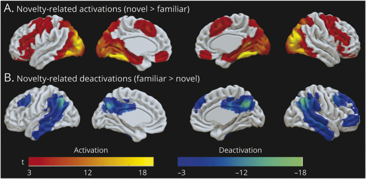
Methods: We studied 499 participants aged 60-88 years from the German Center for Neurodegenerative Diseases Longitudinal Cognitive Impairment and Dementia Study (DELCODE) who underwent task-fMRI. Participants included 163 cognitively normal (healthy control, HC) individuals, 222 SCD, 82 MCI, and 32 patients with clinical diagnosis of mild AD. CSF levels of β-amyloid 42/40 ratio and phosphorylated-tau181 were available from 232 participants. We used region-based analyses to assess novelty-related activity (novel > highly familiar scenes) in entorhinal cortex, hippocampus, and precuneus as well as whole-brain voxel-wise analyses. First, general linear models tested differences in fMRI activity between participant groups. Complementary regression models tested quadratic relationships between memory impairment and activity. Second, relationships of activity with AD CSF biomarkers and brain volume were analyzed. Analyses were controlled for age, sex, study site, and education.

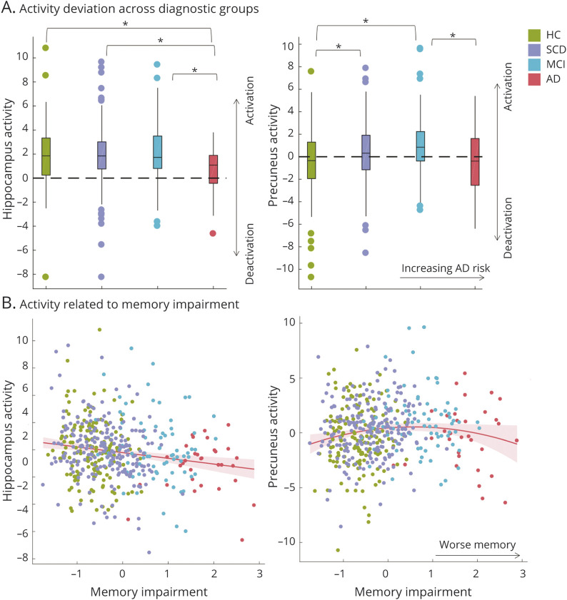
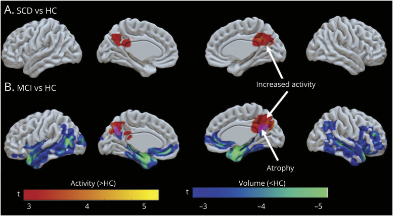
Results: In the precuneus, we observed an inverted U-shaped pattern of novelty-related activity across groups, with higher activity in SCD and MCI compared with HC, but not in patients with AD who showed relatively lower activity than MCI. This nonlinear pattern was confirmed by a quadratic relationship between memory impairment and precuneus activity. Precuneus activity was not related to AD biomarkers or brain volume. In contrast to the precuneus, hippocampal activity was reduced in AD dementia compared with all other groups and related to AD biomarkers.

Discussion: Novelty-related activity in the precuneus follows a nonlinear pattern across the clinical spectrum of increased AD risk. Although the underlying mechanism remains unclear, increased precuneus activity might represent an early signature of memory impairment. Our results highlight the nonlinearity of activity alterations that should be considered in clinical trials using functional outcome measures or targeting hyperactivity.


Figures

Figure 1
Figure 1. Differences in Region-Specific Novelty Activity Between Diagnostic Groups and With Increasing Memory Impairment
(A) Mean fMRI activity (raw betas) for the novelty contrast (novel—familiar scenes) in the hippocampus and precuneus across diagnostic groups. Hippocampal activity was reduced in AD relative to all other groups. Precuneus activity followed an inverted U-shaped pattern with more advanced risk stages for AD. *Significant group differences surviving Bonferroni-Holm correction for the 5 group comparisons of interest (AD < HC < SCD/MCI) with p < 0.05. (B) Activation deviations related to memory performance as a continuous measure of clinical impairment. The memory factor score was inverted (*−1) to represent memory impairment for display purposes. The hippocampus showed a linear but the precuneus a quadratic pattern of activity deviations with increasing memory impairment. AD = Alzheimer disease dementia; HC = healthy control; MCI = mild cognitive impairment; SCD = subjective cognitive decline.
Figure 2
Figure 2. Whole-Brain Voxel-Wise Novelty Activation Pattern in Cognitively Normal Older Adults
(A) Higher fMRI activity for novel than familiar scenes in cognitively normal older adults (N = 163) is seen in a frontal network, the supplementary motor cortex, the medial temporal lobe including the hippocampus and parahippocampal cortex, and the occipital regions bilaterally. (B) Lower fMRI activity for novel compared with familiar scenes (i.e., novelty-related deactivation) is seen in the posterior midline, lateral temporal, temporo-parietal, and frontal regions. Results are depicted at <0.05 (FWE, cluster-level, cluster-forming threshold p = 0.001).
Figure 3
Figure 3. Whole-Brain Voxel-Wise Pattern of Increased Activity and Reduced GM Volume in SCD and MCI
Two-sample t tests revealed higher novelty activity in SCD (A) and MCI (B) relative to cognitively normal older adults in the precuneus (red colors), a region usually deactivating for novel relative to familiar scenes (Figure 2B). fMRI results are depicted at pvoxel <0.005 (uncorrected), pcluster <0.05 FWE corrected. Reduced gray matter volume was seen in the MCI only (B) comprising temporal lobe, frontal regions, and the precuneus (blue colors). Voxel-based morphometry results are depicted p < 0.05 (FWE, cluster-level, cluster-forming threshold p = 0.001). MCI = mild cognitive impairment; SCD = subjective cognitive decline.

References

    1. Putcha D, Brickhouse M, O'Keefe K, et al. . Hippocampal hyperactivation associated with cortical thinning in Alzheimer's disease signature regions in non-demented elderly adults. J Neurosci. 2011;31(48):17680-17688. - PMC - PubMed
    1. Celone KA, Calhoun VD, Dickerson BC, et al. . Alterations in memory networks in mild cognitive impairment and Alzheimer's disease: an independent component analysis. J Neurosci. 2006;26(40):10222-10231. - PMC - PubMed
    1. Kircher TT, Weis S, Freymann K, et al. . Hippocampal activation in patients with mild cognitive impairment is necessary for successful memory encoding. J Neurol Neurosurg Psychiatry. 2007;78(8):812-818. - PMC - PubMed
    1. Machulda MM, Ward HA, Borowski B, et al. . Comparison of memory fMRI response among normal, MCI, and Alzheimer's patients. Neurology. 2003;61(4):500-506. - PMC - PubMed
    1. Sperling RA, LaViolette PS, O'Keefe K, et al. . Amyloid deposition is associated with impaired default network function in older persons without dementia. Neuron. 2009;63(2):178-188. - PMC - PubMed

Publication types

MeSH terms