. 2022 Aug 22;72(1):21.
doi: 10.1186/s12576-022-00846-0.

Hypothalamic orexinergic neurons modulate pain and itch in an opposite way: pain relief and itch exacerbation

Affiliations

Hypothalamic orexinergic neurons modulate pain and itch in an opposite way: pain relief and itch exacerbation

Tatsuroh Kaneko et al. J Physiol Sci. .

Abstract

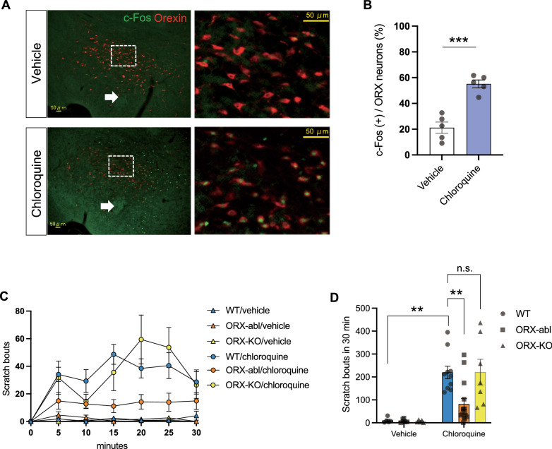
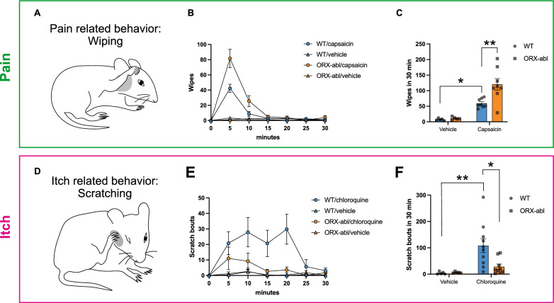
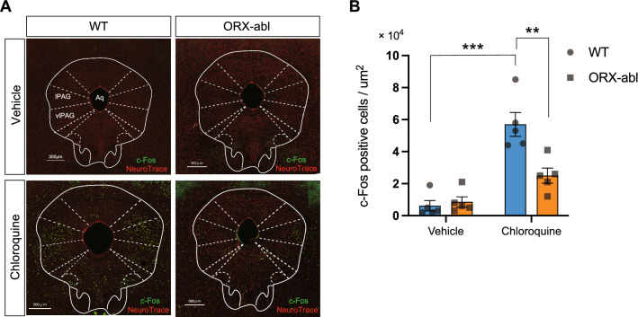
Pain and itch are recognized as antagonistic sensations; pain suppresses itch and inhibition of pain generates itch. There is still a lack of evidence about the neural mechanism of the interaction between pain and itch in the central nervous system. In this study, we focused on the orexin (ORX) neurons in the lateral hypothalamus (LH), which mediate various "defense responses" when animals confront stressors. We found that the scratching behaviors induced by the pruritogen were significantly suppressed in ORX-neuron-ablated (ORX-abl) mice. The exaggerated pain behavior and attenuated itch behavior observed in ORX-abl mice indicated that ORX neurons modulate pain and itch in an opposite way, i.e., pain relief and itch exacerbation. In addition, most of the ORX neurons responded to both pain and itch input. Our results suggest that ORX neurons inversely regulate pain- and itch-related behaviors, which could be understood as a defense response to cope with stress environment.

Keywords: Itch; Lateral hypothalamus; Orexin neuron; Pain.


Conflict of interest statement

The authors declare that they have no competing interests.

Figures

Fig. 1
Fig. 1
Orexin neuron ablation mice, but not orexin peptide-deficient mice, showed the decrease of pruritogen-induced scratching behavior. A c-Fos expression (green) in orexin neurons (red) in the lateral hypothalamus after intradermal injection of vehicle and chloroquine. B The quantification of c-Fos expression showed a significant increase in c-Fos-positive cells in orexin neurons (%) after the chloroquine treatment compared with the vehicle control. n = 5 for each group. C Time course of the number of scratching behaviors induced by intradermal chloroquine injection. The plots indicate the cumulative number of scratching bouts recorded every 5 min. D The scratching behavior within 30 min after chloroquine injection was significantly decreased in ORX-abl mice (n = 15), but not in ORX-KO mice (n = 7), compared with WT mice (n = 10). WT, wild-type mice; ORX-abl, orexin-neuron-ablated mice; ORX-KO, orexin-peptide-deficient mice. The arrows in A indicate fornix. The white boxed area in A left is magnified in A right. The data represent the mean ± SEM. ***P < 0.001 (unpaired t-test). **P < 0.01 (two-way ANOVA with post hoc Tukey’s multiple comparisons test)
Fig. 2
Fig. 2
Orexin neurons modulate pain and itch sensations in an opposite way. A Illustration of the wiping behavior using a single forelimb (pain-related behavior). B Time course of the number of wiping behaviors induced by capsaicin injection into the cheek. The plots indicate the cumulative number of wiping events recorded every 5 min. C The number of wiping events within 30 min after capsaicin injection was significantly increased in ORX-abl mice (n = 8) compared with WT mice (n = 8). D Illustration of the scratching behavior using the hind leg (itch-related behavior). E Time course of the number of scratching bouts induced by chloroquine injection into the right cheek. The plots indicate the cumulative number of scratching bouts recorded every 5 min. F The number of scratching bouts within 30 min after chloroquine injection was significantly decreased in ORX-abl mice (n = 9) compared with WT mice (n = 10). The data represent the mean ± SEM. *P < 0.05, **P < 0.01 (two-way ANOVA with post hoc Tukey’s multiple comparisons test)
Fig. 3
Fig. 3
The same orexin neuron responded to both itch and pain stimulation. A Schematic diagram representing the temporally controlled expression of the GCaMP6 protein under c-Fos promoter using the Tet-off system in cFos-tTA; TetO-GCaMP6 mice. B Diagram showing the experimental procedure, doxycycline timing, and schedule. C Expression of GCaMP6 (green) and c-Fos (blue) in orexin neurons (red) after the injection of vehicle or stimulants (capsaicin and chloroquine). Yellow arrowheads indicate the ORX cells overlapping with GCaMP6 and c-Fos expression. D The quantification of GCaMP6 and c-Fos expression in orexin neurons revealed a significant increase of GCaMP6 and c-Fos double-positive cells in orexin neurons (%) after pain and itch stimulation compared with the vehicle control. n = 5 for each group. The data represent the mean ± SEM. ***P < 0.001 (unpaired t-test). E Venn diagram showing the response ratio of orexin neurons to pain and itch stimulation. Left, the proportion of orexin neurons that responded to vehicle injection. Right, the proportion of orexin neurons that responded to pain (GCaMP6; green) and itch (c-Fos; red) stimulation. More than half of the orexin neurons responded to both pain and itch stimulation
Fig. 4
Fig. 4
Attenuation of the pruritogen-induced activation of PAG neurons in orexin-neuron-ablated mice. A c-Fos expression (green) in lPAG/vlPAG neurons (red) after intradermal injection of vehicle or chloroquine into WT and ORX-abl mice. B The densities of c-Fos-positive neurons in the lPAG/vlPAG area after intradermal administration of vehicle or chloroquine were plotted. The c-Fos expression level after chloroquine injection was significantly decreased in ORX-abl mice compared with that in WT mice. The data represent the mean ± SEM. **P < 0.01, ***P < 0.001 (two-way ANOVA with post hoc Tukey’s multiple comparison test), n = 5 for all groups

References

    1. Schmelz M. Itch and pain. Neurosci Biobehav Rev. 2010;34:171–176. doi: 10.1016/j.neubiorev.2008.12.004. - DOI - PubMed
    1. Davidson S, Zhang X, Khasabov SG, Simone DA, Giesler GJ. Relief of itch by scratching: state-dependent inhibition of primate spinothalamic tract neurons. Nat Neurosci. 2009;12:544–546. doi: 10.1038/nn.2292. - DOI - PMC - PubMed
    1. Ikoma A, Rukwied R, Ständer S, Steinhoff M, Miyachi Y, Schmelz M. Neurophysiology of pruritus: interaction of itch and pain. Arch Dermatol. 2003;139:1475–1478. doi: 10.1001/archderm.139.11.1475. - DOI - PubMed
    1. Dong X, Dong X. Peripheral and central mechanisms of itch. Neuron. 2018;98:482–494. doi: 10.1016/j.neuron.2018.03.023. - DOI - PMC - PubMed
    1. Han L, Ma C, Liu Q, Weng HJ, Cui Y, Tang Z, Kim Y, Nie H, Qu L, Patel KN, Li Z, McNeil B, He S, Guan Y, Xiao B, Lamotte RH, Dong X. A subpopulation of nociceptors specifically linked to itch. Nat Neurosci. 2013;16:174–182. doi: 10.1038/nn.3289. - DOI - PMC - PubMed