. 2022 Aug 17;2(4):419-436.
doi: 10.1021/acsbiomedchemau.2c00007. Epub 2022 Apr 8.

Synergetic Antimicrobial Activity and Mechanism of Clotrimazole-Linked CO-Releasing Molecules

Affiliations

Synergetic Antimicrobial Activity and Mechanism of Clotrimazole-Linked CO-Releasing Molecules

Sofia S Mendes et al. ACS Bio Med Chem Au. .

Abstract

Several metal-based carbon monoxide-releasing molecules (CORMs) are active CO donors with established antibacterial activity. Among them, CORM conjugates with azole antibiotics of type [Mn(CO)3(2,2'-bipyridyl)(azole)]+ display important synergies against several microbes. We carried out a structure-activity relationship study based upon the lead structure of [Mn(CO)3(Bpy)(Ctz)]+ by producing clotrimazole (Ctz) conjugates with varying metal and ligands. We concluded that the nature of the bidentate ligand strongly influences the bactericidal activity, with the substitution of bipyridyl by small bicyclic ligands leading to highly active clotrimazole conjugates. On the contrary, the metal did not influence the activity. We found that conjugate [Re(CO)3(Bpy)(Ctz)]+ is more than the sum of its parts: while precursor [Re(CO)3(Bpy)Br] has no antibacterial activity and clotrimazole shows only moderate minimal inhibitory concentrations, the potency of [Re(CO)3(Bpy)(Ctz)]+ is one order of magnitude higher than that of clotrimazole, and the spectrum of bacterial target species includes Gram-positive and Gram-negative bacteria. The addition of [Re(CO)3(Bpy)(Ctz)]+ to Staphylococcus aureus causes a general impact on the membrane topology, has inhibitory effects on peptidoglycan biosynthesis, and affects energy functions. The mechanism of action of this kind of CORM conjugates involves a sequence of events initiated by membrane insertion, followed by membrane disorganization, inhibition of peptidoglycan synthesis, CO release, and break down of the membrane potential. These results suggest that conjugation of CORMs to known antibiotics may produce useful structures with synergistic effects that increase the conjugate's activity relative to that of the antibiotic alone.


Conflict of interest statement

The authors declare no competing financial interest.

Figures
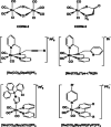
Figure 1
Figure 1
Selected CORMs with antimicrobial activity.
Figure 2
Figure 2
Conjugates prepared by combining the fac-{M(CO)3}d6 fragment with bidentate ligands and clotrimazole.
Scheme 1
Scheme 1. Synthesis of the Clotrimazole–CORM Conjugates
M = Mn and Re; X = Br and I; OTf = O3SCF3 (triflate); L–L = Bpy, Biq, PyBzim, Bpydinon, PyNHC, and bisNHC; and Ctz = clotrimazole. OTf (O3SCF3, triflate).
Figure 3
Figure 3
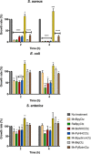
S. aureus cells are sensitive to conjugated CORMs. Cells of S. aureus MRSA, E. coli MG1655, and S. enterica SL1344 were treated with the indicated CORMs (35 μM for Gram-negative bacteria and 10 μM for S. aureus). The percentage of the growth rate was determined in relation to untreated cells collected at 2 h and 4 h. Data represent the average of three independent biological samples, with error bars representing the standard deviation. *P < 0.05, **P < 0.01, ***P < 0.001, and ****P < 0.0001.
Figure 4
Figure 4
Release of CO from conjugated CORMs into blood and bacteria. (A) Percentage of CO–hemoglobin formed in blood samples incubated, for 1 h, with MnBpyCtz, ReBpyCtz, MnbisNHCCtz, MnPyNHCCtz, MnBiqCtz, MnPyBzimCtz, and DMSO. (B) Percentage of Met–hemoglobin formed in blood samples incubated for 1 h with MnBpyCtz, ReBpyCtz, MnbisNHCCtz, MnPyNHCCtz, MnBiqCtz, MnPyBzimCtz, and DMSO. (C) Percentage of CO–hemoglobin bound in blood samples incubated, for 30 min, with ALF21. (D) Percentage of Met–hemoglobin bound in blood samples incubated, for 30 min, with ALF21. (E) Fluorescence microscopy images of S. aureus MRSA CORM–Ctz conjugates (10 μM, 15 min) and incubated with the fluorescent probe COP-1. Representative images of fluorescent cells treated with MnBpyCtz (a), ReBpyCtz (b), MnbisNHCCtz (c), MnPyNHCCtz (d), MnBiqCtz (e), and MnPyBzimCtz (f) (upper row) and their corresponding bright field images (g, h, i, j, k, and l, respectively) (lower images). The population values correspondent to fluorescent green cells represented on the images are 80% (a), 74% (b), 84% (c), 44% (c), 86% (d) and 85% (e), respectively. At least 300 cells, from at least three independent experiments, were analyzed for each condition.
Figure 5
Figure 5
Effect of ReBpyCtz on the cell envelope of growing B. subtilis and S. aureus cells. (A) Firefly luciferase bioreporter profiling demonstrates liaI promotor induction by ReBpyCtz. The liaI promotor responds to cell envelope damage and particularly to agents interfering with cycling of the membrane-standing undecaprenyl-P precursor. Luminescence values recorded at a single predetermined time point after ReBpyCtz addition (see the Material and Methods section) were normalized to the untreated control (100%, black, horizontal dashed line) and plotted against the concentration of ReBpyCtz. At the MIC, the liaI bioreporter is induced, while the signals for the noninduced bioreporters fall below the background level, indicating impaired biosynthetic capacity. The corresponding experiments for Ctz, ReBpyBr, and reference antibiotics are shown in Supporting Information Figure S2. Error bars represent the SD of two biological replicates (i.e., cultures grown and treated independently on different days) with two technical replicates per test day. (B) ReBpyCtz and Ctz disturb the topology of the cytoplasmic membrane. Membrane staining by FM 5-95 of S. aureus NCTC 8325 treated for 30 min with Ctz (8 μg/mL, 2× MIC), ReBpyBr (64 μg/mL), and ReBpyCtz (0.5 μg/mL, 2× MIC) compared to the negative control treated with DMSO. Scale bar, 1 μm. One experiment representative of three biological replicates is shown. (C) Time-resolved effect of Ctz and ReBpyCtz on the membrane potential of S. aureus NCTC8325 measured by DiOC2 (3) staining in relation to the respective MICs. Protonophore CCCP (5 μM, 0.4× MIC) was used as a positive control and DMSO as a negative control. The arrow indicates the time point of compound addition. Four biological replicates and SD are presented. (D) Membrane integrity is not severely affected. Exposure of S. aureus NCTC8325 to either Ctz (16 μg/mL, 4× MIC), ReBpyCtz (1 μg/mL, 4× MIC), or ReBpyBr (64 μg/mL) for 120 min, followed by staining with membrane-permeant Syto9 (green) and membrane impermeant PI (red). Nisin (100 μg/mL) served as a positive control and DMSO (1%) as a negative control. Merged overlay of three photographs of the same cells acquired in the brightfield, green, or red fluorescence channel. Scale bar, 2 μm. (E) ReBpyBr leads to membrane blebbing in B. subtilis 168, indicative of peptidoglycan weakening. Bright-field visualization of the formation of blebs after 30 min treatment with ReBpyBr (64 μg/mL) or vancomycin (Van, positive control, 2 μg/mL). No blebs were induced by Ctz (8 μg/mL, 4× MIC), ReBpyCtz (1 μg/mL, 4× MIC), or DMSO (1%, negative control). Scale bar, 2 μm. (F) UDP-MurNAc-pentapeptide accumulation in S. aureus ATCC 29213 after 30 min of treatment with DMSO (negative control), vancomycin (2 μg/mL, 8× MIC, positive control), Ctz (40 μg/mL, 10× MIC), ReBpyBr (64 μg/mL), or ReBpyCtz (0.5, 2.5, and 5 μg/mL, corresponding to 2×, 10×, and 20× MIC, respectively) shown as AUC values of EICs. An exemplary EIC (shown for the ReBpyCtz at 10× MIC sample) is presented as an inset, depicting the mass of UDP-MurNAc-pentapeptide (m/z = 1148.354) measured in the negative ionization mode. Further EICs and corresponding total ion chromatograms are presented in Supporting Information Figure S5. (G) Monitoring nascent peptidoglycan in growing B. subtilis cells. HADA labeling of B. subtilis 168 after 5 min exposure to Ctz (4 μg/mL, 2× MIC), ReBpyCtz (0.5 μg/mL, 2× MIC or 1 μg/mL, 4× MIC), or CCCP (5 μM) compared to DMSO (0.5%), the negative control. Fluorescence channel (bottom) and overlay with the phase contrast (top). Microscopic settings were selected to yield a good signal in the DMSO control and then kept constant for all micrographs. Scale bar, 5 μm. (H) Quantification of (G). The violin plots represent the relative septal HADA intensities quantified from ≥100 cells per experiment from three independent biological replicates; ****P < 0.0001.
Figure 6
Figure 6
Inhibition of in vitro peptidoglycan synthesis by ReBpyCtz and interaction with peptidoglycan precursors. (A) Schematic of lipid II synthesis in S. aureus NCTC8325. UDP-MurNAc-PP and lipid carrier C55P are used as substrates for lipid I synthesis by MraY. MurG catalyzes the addition of GlcNAc from UDP-GlcNAc, yielding lipid II. (B) Influence of ReBpyCtz, ReBpyBr, and Ctz on lipid II synthesis using MraY- and MurG-containing membrane preparations of Micrococcus luteus. At a 10-fold molar excess over C55P, ReBpyCtz and Ctz slightly reduced the amount of lipid II formed, while ReBpyBr did not. NC, negative control, reaction mixture without membranes; DMSO (1%). Error bars represent the SD of two replicates. (C) Influence of ReBpyCtz on lipid II synthesis from purified lipid I (2 nmol) and UDP-GlcNAc by purified MurG. Test compounds were applied in DMSO (1%) at a 10-fold molar excess over lipid I. TLC curves showing the extracted lipids at the end of the reaction. Vancomycin was used as an inhibition control; its binding to lipid I retains the complex in the aqueous phase and prevents it from being extracted. ReBpyCtz sightly inhibited the conversion of lipid I to lipid II. (D) Quantification of the bands visible on the TLC plate in (C). ReBpyCtz inhibits the MurG reaction. Error bars represent the SD of two replicates. (E) LiaI lux bioreporter strain based on the Photorhabdus luminescens luciferase system, yielding a continuous fluorescence signal. The liaI promotor in B. subtilis 168 is induced upon treatment with ReBpyCtz and Ctz around their respective MIC levels, albeit to different extents. No induction was observed for ReBpyBr. Vancomycin (2 μg/mL, 2× MIC) served as a positive control. (F,G) Antagonization of the bioreporter induction by peptidoglycan precursor addition. In the case of ReBpyCtz (F), purified lipid II (triangles) antagonized the most effectively, and in the case of clotrimazole (G), C55PP antagonized the best. C55P was not significantly effective. The depicted experiment is representative of two independent biological replicates.

Similar articles

Cited by

References

    1. Motterlini R.; Otterbein L. E. The Therapeutic Potential of Carbon Monoxide. Nat. Rev. Drug Discovery 2010, 9, 728–743. 10.1038/nrd3228. - DOI - PubMed
    1. Nobre L. S.; Seixas J. D.; Romão C. C.; Saraiva L. M. Antimicrobial Action of Carbon Monoxide-Releasing Compounds. Antimicrob. Agents Chemother. 2007, 51, 4303–4307. 10.1128/aac.00802-07. - DOI - PMC - PubMed
    1. Mendes S. S.; Miranda V.; Saraiva L. M. Hydrogen Sulfide and Carbon Monoxide Tolerance in Bacteria. Antioxidants 2021, 10, 729–746. 10.3390/antiox10050729. - DOI - PMC - PubMed
    1. Wareham L. K.; Poole R. K.; Tinajero-Trejo M. CO-Releasing Metal Carbonyl Compounds as Antimicrobial Agents in the Post-Antibiotic Era. J. Biol. Chem. 2015, 290, 18999–19007. 10.1074/jbc.r115.642926. - DOI - PMC - PubMed
    1. Johnson T. R.; Mann B. E.; Teasdale I. P.; Adams H.; Foresti R.; Green C. J.; Motterlini R. Metal Carbonyls as Pharmaceuticals? [Ru(CO)3Cl(Glycinate)], a CO-Releasing Molecule with an Extensive Aqueous Solution Chemistry. Dalton Trans. 2007, 15, 1500–1508. 10.1039/b613629j. - DOI - PubMed

LinkOut - more resources