

Extracellular Volume Fraction Based on Cardiac Magnetic Resonance T1 Mapping: An Effective Way to Evaluate Cardiac Injury Caused by Cardiac Amyloidosis in Patients with Multiple Myeloma
- PMID: 35996622
- PMCID: PMC9392639
- DOI: 10.1155/2022/3094933
Extracellular Volume Fraction Based on Cardiac Magnetic Resonance T1 Mapping: An Effective Way to Evaluate Cardiac Injury Caused by Cardiac Amyloidosis in Patients with Multiple Myeloma
Abstract
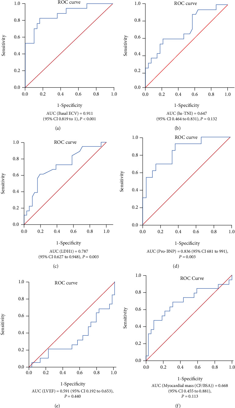
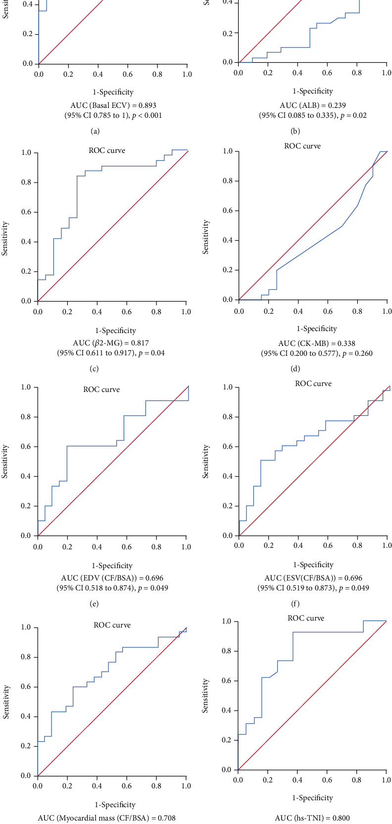
Multiple myeloma (MM) is a hematological malignancy of plasma cell origin. Cardiac amyloidosis (CA) is a common form of heart damage caused by MM and is associated with a poor prognosis. This study was a prospective cohort study and was aimed at evaluating the clinical predictive value of extracellular volume fraction (ECV) based on cardiovascular magnetic resonance (CMR) T1 mapping for cardiac amyloidosis and cardiac dysfunction in MM patients. Fifty-one newly diagnosed MM patients in Zhongnan Hospital of Wuhan University were enrolled in the study. A total of 19 patients (19/51; 37.25%) developed CA. The basal ECV of CA group was significantly higher than that of the non-CA group (p < 0.01). Multivariate logistic regression analysis showed that basal ECV (OR = 1.551, 95% CI 1.084-2.219, p < 0.05) and LDH1 level (OR = 1.150, 95% CI 1.010-1.310, p < 0.05) were two independent risk factors for CA. Further study demonstrated that basal ECV in the heart failure group was significantly higher than that of the nonheart failure group (p < 0.01). Notably, ROC curve showed that basal ECV had a good predictive value for CA and heart failure, with AUC of 0.911 and 0.893 (all p < 0.01), and the best cutoff values of 38.35 and 37.45, respectively. Taken together, basal ECV is a good predictor of CA and heart failure for MM patients.
Copyright © 2022 Minghui Liu et al.
Conflict of interest statement
The authors have no conflict of interest.
Figures


Similar articles
-
Prognostic value of heart failure in hemodialysis-dependent end-stage renal disease patients with myocardial fibrosis quantification by extracellular volume on cardiac magnetic resonance imaging.BMC Cardiovasc Disord. 2020 Jan 10;20(1):12. doi: 10.1186/s12872-019-01313-2. BMC Cardiovasc Disord. 2020. Retraction in: BMC Cardiovasc Disord. 2020 Sep 15;20(1):407. doi: 10.1186/s12872-020-01688-7. PMID: 31924159 Free PMC article. Retracted.
-
Regional Amyloid Burden Differences Evaluated Using Quantitative Cardiac MRI in Patients with Cardiac Amyloidosis.Korean J Radiol. 2021 Jun;22(6):880-889. doi: 10.3348/kjr.2020.0579. Epub 2021 Feb 24. Korean J Radiol. 2021. PMID: 33686816 Free PMC article.
-
Diagnostic value of the novel CMR parameter "myocardial transit-time" (MyoTT) for the assessment of microvascular changes in cardiac amyloidosis and hypertrophic cardiomyopathy.Clin Res Cardiol. 2021 Jan;110(1):136-145. doi: 10.1007/s00392-020-01661-6. Epub 2020 May 5. Clin Res Cardiol. 2021. PMID: 32372287 Free PMC article.
-
Diagnostic value of cardiovascular magnetic resonance in comparison to endomyocardial biopsy in cardiac amyloidosis: a multi-centre study.Clin Res Cardiol. 2021 Apr;110(4):555-568. doi: 10.1007/s00392-020-01771-1. Epub 2020 Nov 10. Clin Res Cardiol. 2021. PMID: 33170349 Free PMC article.
-
Myocardial structural and functional changes in cardiac amyloidosis: insights from a prospective observational patient registry.Eur Heart J Cardiovasc Imaging. 2023 Dec 21;25(1):95-104. doi: 10.1093/ehjci/jead188. Eur Heart J Cardiovasc Imaging. 2023. PMID: 37549339 Free PMC article.
Cited by
-
T1 mapping in evaluation of clinicopathologic factors for rectal adenocarcinoma.Abdom Radiol (NY). 2024 Jan;49(1):279-287. doi: 10.1007/s00261-023-04045-2. Epub 2023 Oct 15. Abdom Radiol (NY). 2024. PMID: 37839066
References
-
- Cooper L. T., Baughman K. L., Feldman A. M., et al. The role of endomyocardial biopsy in the management of cardiovascular disease: a scientific statement from the American Heart Association, the American College of Cardiology, and the European Society of Cardiology. Circulation . 2007;116(19):2216–2233. doi: 10.1161/CIRCULATIONAHA.107.186093. - DOI - PubMed
MeSH terms
LinkOut - more resources
Full Text Sources
Medical
