. 2022 Aug 12;12(5):20220017.
doi: 10.1098/rsfs.2022.0017. eCollection 2022 Oct 6.

Potential of stem cell seeded three-dimensional scaffold for regeneration of full-thickness skin wounds

Affiliations

Potential of stem cell seeded three-dimensional scaffold for regeneration of full-thickness skin wounds

Irfan Khan et al. Interface Focus. .

Abstract

Hypoxic wounds are tough to heal and are associated with chronicity, causing major healthcare burden. Available treatment options offer only limited success for accelerated and scarless healing. Traditional skin substitutes are widely used to improve wound healing, however, they lack proper vascularization. Mesenchymal stem cells (MSCs) offer improved wound healing; however, their poor retention, survival and adherence at the wound site negatively affect their therapeutic potential. The aim of this study is to enhance skin regeneration in a rat model of full-thickness dermal wound by transplanting genetically modified MSCs seeded on a three-dimensional collagen scaffold. Rat bone marrow MSCs were efficiently incorporated in the acellular collagen scaffold. Skin tissues with transplanted subcutaneous scaffolds were histologically analysed, while angiogenesis was assessed both at gene and protein levels. Our findings demonstrated that three-dimensional collagen scaffolds play a potential role in the survival and adherence of stem cells at the wound site, while modification of MSCs with jagged one gene provides a conducive environment for wound regeneration with improved proliferation, reduced inflammation and enhanced vasculogenesis. The results of this study represent an advanced targeted approach having the potential to be translated in clinical settings for targeted personalized therapy.

Keywords: biomaterials; hypoxia; mesenchymal stem cells; tissue engineering; transfection; wound regeneration.


Figures

Figure 1.
Figure 1.
Characterization of MSCs: immunofluorescent micrographs showing MSC markers CD44, CD90, CD29 and CD117, haematopoietic marker CD45, and control MSCs stained only with Alexa fluor 546 goat anti-mouse secondary antibody. Nuclei were stained with DAPI.
Figure 2.
Figure 2.
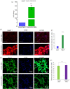
Jagged 1 expression in MSCs. (a) Gene expression of jagged 1 in normal and transfected MSCs. (b) Protein expression of jagged 1 in normal and transfected MSCs. (c) Quantification of fluorescent intensity for jagged 1 by ImageJ. (d) Immunostaining for MSC stemness marker stro-1. (e) Quantification of fluorescent intensity for stro-1 by ImageJ. Data are presented as mean ± s.e.m. (n = 3) with significance level ***p < 0.001.
Figure 3.
Figure 3.
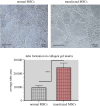
In vitro tube formation assay: normal and transfected MSCs showing tube formation in Matrigel assay. Number of tubes were counted from three biological replicates and plotted. Data are presented as mean ± s.e.m. (n = 3) with significance level ***p < 0.001.
Figure 4.
Figure 4.
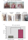
Hypoxic wound model. (a) Hypoxic wound model was developed on the dorsal surface of rat skin. (b) Gross macroscopic examination of wound tissues at Day 14. (c) Gene expression analysis of HIF-1α was performed by qRT-PCR to confirm that the wound is of hypoxic nature. Data are presented as mean ± s.e.m. (n = 3) with significance level *p < 0.05.
Figure 5.
Figure 5.
Gene expression analysis: bar graphs showing the expression levels of cell survival and proliferation, anti-inflammatory, and angiogenesis genes in normal skin, wound model, collagen scaffold only transplanted group, and groups transplanted with normal MSC and jagged 1 transfected MSC seeded scaffolds. For statistical analysis, one-way ANOVA was performed, followed by the Bonferroni post hoc test. Data are presented as mean ± s.e.m. (n = 3). *p < 0.05, **p < 0.01 and ***p < 0.001.
Figure 6.
Figure 6.
Histological analysis of wound tissue sections: H&E and Masson's trichrome staining showing tissue architecture after 14 days of wound induction and transplantation. Jagged 1 transfected MSC seeded scaffold group showed complete re-epithelialization with collagen ECM, vascularization and remodelling of the tissue framework with a distinct layer of epidermis and dermis without scar.
Figure 7.
Figure 7.
Immunohistochemical staining for α-SMA and VEGF. (a) Representative images of harvested tissue from normal, scaffold only, MSC seeded scaffold and jagged 1 transfected MSC seeded scaffold groups showing enhanced angiogenesis and VEGF expression. (b,c) Quantification of fluorescent intensity for α-SMA and VEGF by ImageJ. (d) Quantitative analysis of blood vessels in normal, scaffold only, MSC seeded scaffold, and jagged 1 transfected MSC seeded scaffold groups. Data are presented as mean ± s.e.m. (n = 3) with significance level ***p < 0.001.

References

    1. Thapa RK, Margolis DJ, Kiick KL, Sullivan MO. 2020. Enhanced wound healing via collagen-turnover-driven transfer of PDGF-BB gene in a murine wound model. ACS App. Bio Mater. 3, 3500-3517. (10.1021/acsabm.9b01147) - DOI - PMC - PubMed
    1. Nourian DA, Mirahmadi BF, Chehelgerdi M, Raeisi DS. 2019. Skin tissue engineering: wound healing based on stem-cell-based therapeutic strategies stem. Cell Res. Ther. 10, 111-120. (10.1186/s13287-019-1212-2) - DOI - PMC - PubMed
    1. Riha SM, Maarof M, Fauzi MB. 2021. Synergistic effect of biomaterial and stem cell for skin tissue engineering in cutaneous wound healing: a concise review. Polymers 13, 1546-1559. (10.3390/polym13101546) - DOI - PMC - PubMed
    1. Kosaric N, Kiwanuka H, Gurtner GC. 2019. Stem cell therapies for wound healing. Exp. Opin. Biol. Ther. 19, 575-585. (10.1080/14712598.2019.1596257) - DOI - PubMed
    1. Ruthenborg RJ, Ban JJ, Wazir A, Takeda N, Kim JW. 2014. Regulation of wound healing and fibrosis by hypoxia and hypoxia-inducible factor-1. Mol. Cells 37, 637-643. (10.14348/molcells.2014.0150) - DOI - PMC - PubMed

LinkOut - more resources