Review
. 2022 Dec 5;23(23):e202200341.
doi: 10.1002/cbic.202200341. Epub 2022 Sep 15.

Epipolythiodioxopiperazine-Based Natural Products: Building Blocks, Biosynthesis and Biological Activities

Affiliations
Review

Epipolythiodioxopiperazine-Based Natural Products: Building Blocks, Biosynthesis and Biological Activities

Eva M Huber. Chembiochem. .

Abstract

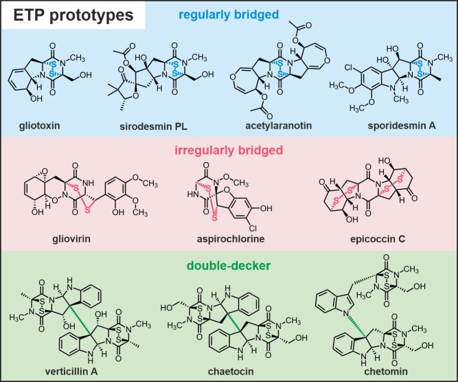
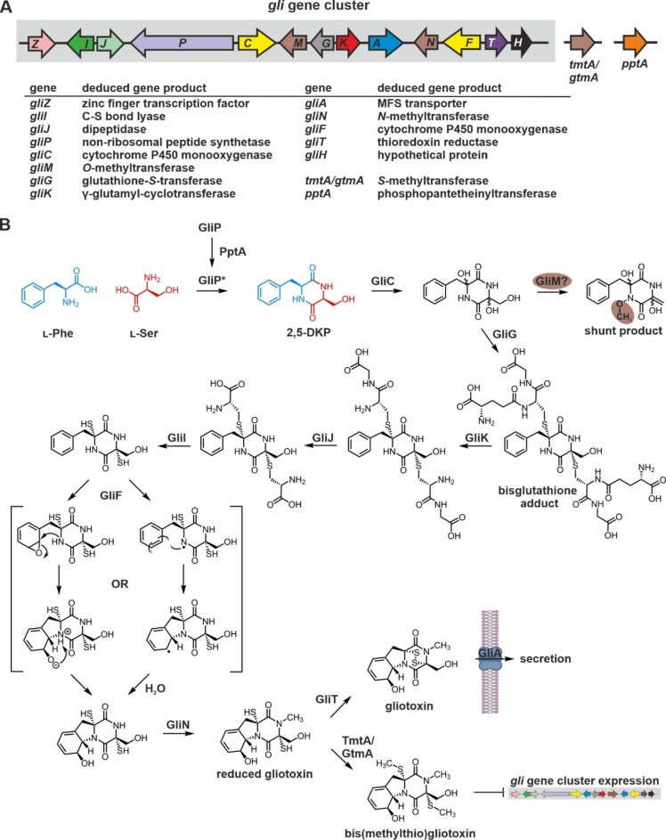
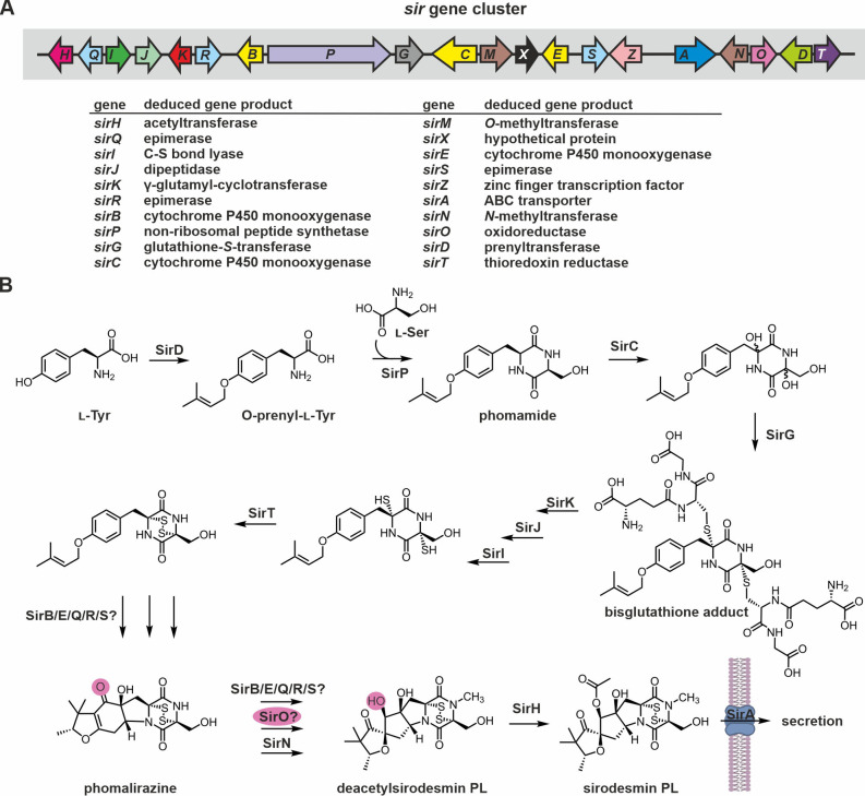
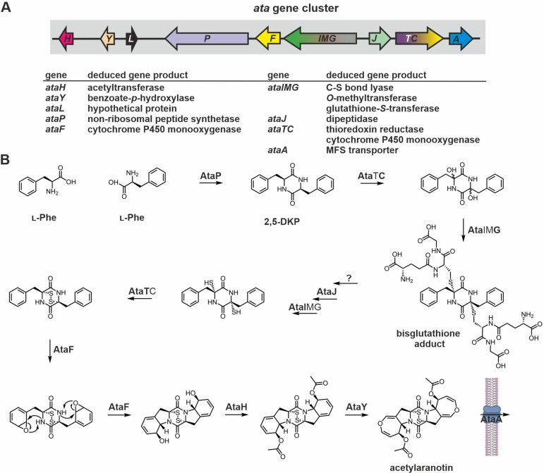
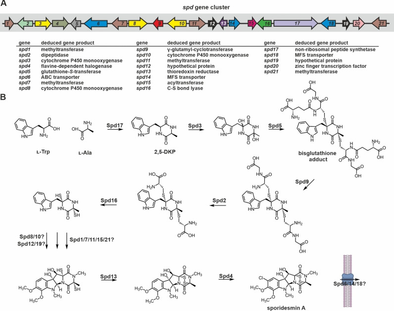
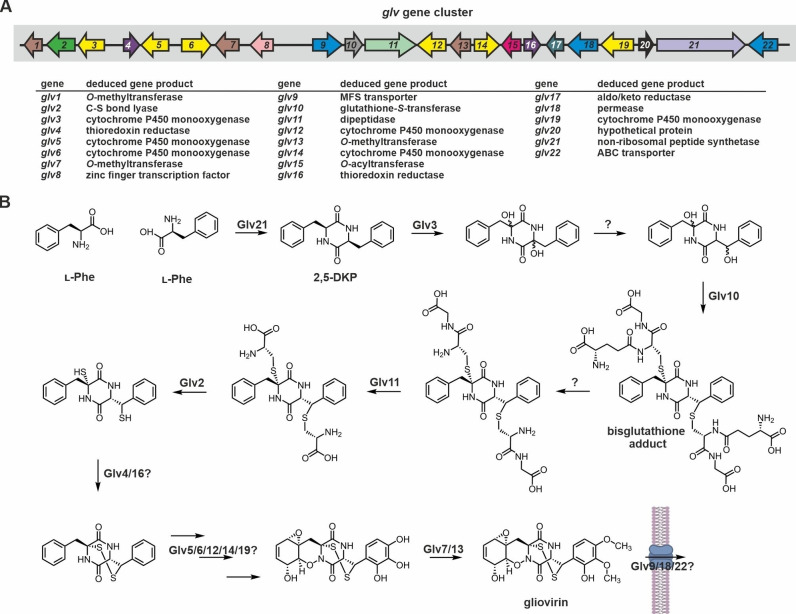
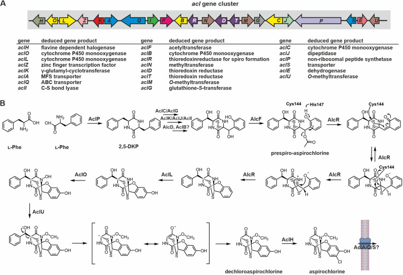
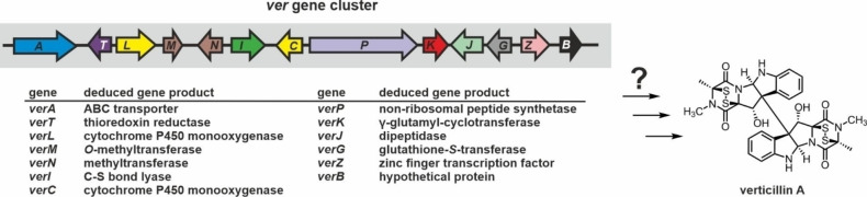
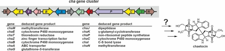
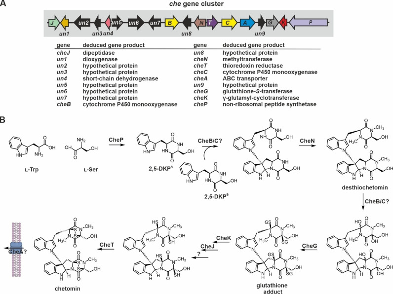
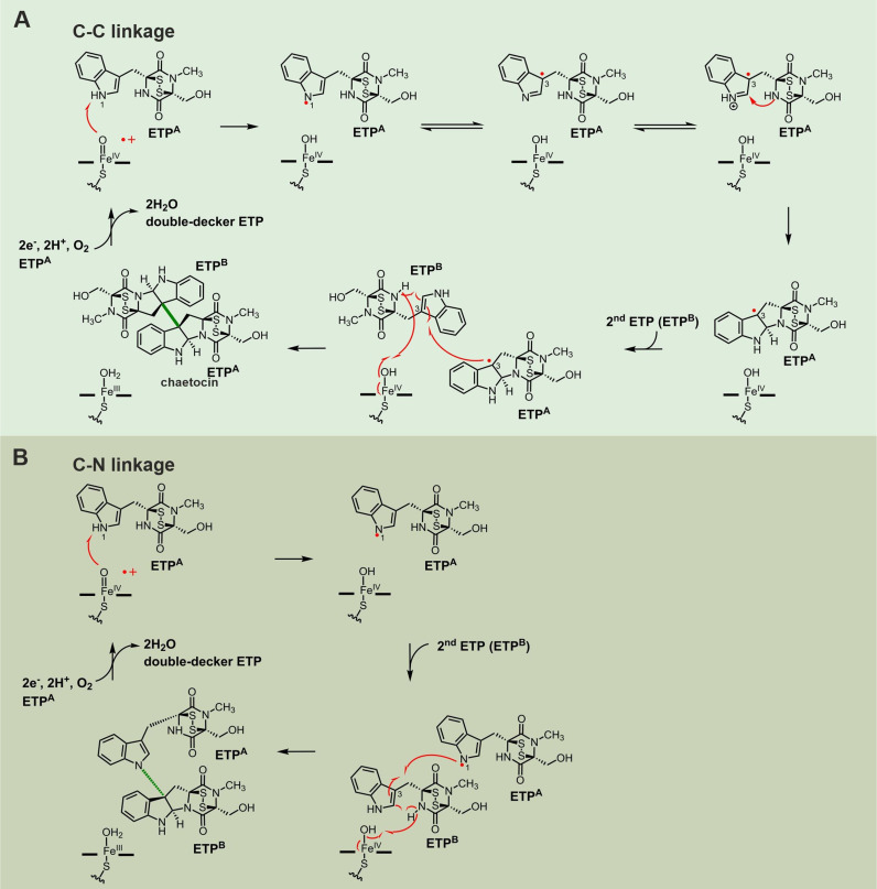
Epipolythiodioxopiperazines (ETPs) are fungal secondary metabolites that share a 2,5-diketopiperazine scaffold built from two amino acids and bridged by a sulfide moiety. Modifications of the core and the amino acid side chains, for example by methylations, acetylations, hydroxylations, prenylations, halogenations, cyclizations, and truncations create the structural diversity of ETPs and contribute to their biological activity. However, the key feature responsible for the bioactivities of ETPs is their sulfide moiety. Over the last years, combinations of genome mining, reverse genetics, metabolomics, biochemistry, and structural biology deciphered principles of ETP production. Sulfurization via glutathione and uncovering of the thiols followed by either oxidation or methylation crystallized as fundamental steps that impact expression of the biosynthesis cluster, toxicity and secretion of the metabolite as well as self-tolerance of the producer. This article showcases structure and activity of prototype ETPs such as gliotoxin and discusses the current knowledge on the biosynthesis routes of these exceptional natural products.

Keywords: biosynthetic gene clusters; disulfide bridges; enzymatic reactions; fungi; toxins.


Conflict of interest statement

The authors declare no conflict of interest.

Figures

Figure 1
Figure 1
Chemical structures and names of prominent epipolythiodioxopiperazine (ETP) compounds. Prototype ETPs are grouped according to the type of their sulfur bridge and their higher‐order structure. In regularly bridged ETPs the disulfide moiety links the two Cα atoms of the 2,5‐diketopiperazine (2,5‐DKP) backbone. Irregularly bridged ETPs are characterized by deviations from this Cα–Cα connection and can be grouped according to the number of cycles their sulfur bridge spans. Sulfur linkages of the Cα–Cβ type only cross the DKP core (gliovirin and aspirochlorine), while epicoccin C presents with two bis‐cross‐ring bridges. In addition, dimeric ETP structures are known. They are classified according to the type of bond connecting their two units: C−C type for verticillin A and chaetocin versus C−N type for chetomin.
Figure 2
Figure 2
Genetic elements required for gliotoxin production and current biosynthesis scheme. (A) Schematic view of the gliotoxin (gli) biosynthesis gene cluster from A. fumigatus.[ 66b , 71 ] Genes are illustrated as colored and labelled arrows. Black ones have not yet been assigned a specific function. Open reading frames within the gli locus are shaded against a gray background (left), while those outside the biosynthetic gene cluster (right) are not. Verified or predicted functions of gene products are listed. (B) Current reaction scheme for the biosynthesis of gliotoxin. The NRPS GliP is activated by the phosphopantetheinyltransferase PptA encoded outside the gli gene cluster and fuses the two starter amino acids l‐Phe (blue) and l‐Ser (red) to the 2,5‐DKP scaffold. The reaction steps until the free dithiol precursor are confirmed and fix in sequence. GliM might act as an O‐methyltransferase on a transient, yet unknown intermediate or on a shunt metabolite as indicated. The order of modifications catalyzed by GliF, GliN and GliT appears to be interchangeable. The reaction trajectory of GliF is unknown, but two potential mechanisms have been suggested. [75b] Disulfide‐bridged gliotoxin is exported outside the fungal cell by a transport protein, while bis‐thiomethylation of reduced gliotoxin by the S‐methyltransferase TmtA (also known as GtmA) encoded outside the gli gene cluster depletes oxidized gliotoxin and thereby serves to dampen expression of gliotoxin biosynthesis genes and ultimately ETP production.[ 75d , 76 ] For details see section 2.1.1.2.
Figure 3
Figure 3
Gene cluster and current biosynthesis scheme for sirodesmin PL production. (A) Schematic view of the sir genes forming the sirodesmin biosynthesis gene cluster in L. maculans.[ 72 , 135 ] Genes are colored according to Figure 2A. (B) Proposed reaction sequence leading to sirodesmin PL. Prominent intermediates are labelled. Putative biosynthesis intermediates produced by SirK and SirJ are not shown, as processing of the glutathione moieties likely proceeds as in gliotoxin biosynthesis. The reactions catalyzed by SirD, SirP, SirC, SirG, SirK, SirJ and SirI are likely fixed in their order. SirO might reduce the ketone group outside the 2,5‐DKP ring of phomalirazine. For details see section 2.1.2.2.
Figure 4
Figure 4
Gene cluster and current biosynthesis scheme for acetylaranotin production. (A) Schematic view of the ata genes in the acetylaranotin biosynthesis gene cluster from A. terreus. [94a] Genes are colored according to Figure 2A. The cluster contains two modular genes that might encode for multifunctional enzymes: AtaTC and AtaIMG. (B) Reaction sequence proposed for the enzymatic production of acetylaranotin – a centrosymmetric ETP. Domains of predicted multifunctional enzymes required for a certain reaction are printed bold. The putative biosynthesis intermediate produced by AtaJ is not shown, as processing of the glutathione moieties likely proceeds as in gliotoxin biosynthesis. The order of reactions catalyzed by the enzymes AtaH and AtaY is interchangeable. For details see section 2.1.3.
Figure 5
Figure 5
Gene cluster and putative biosynthesis scheme for sporidesmin A production. (A) Schematic view of the predicted sporidesmin A (spd) biosynthesis gene cluster from P. chartarum according to a preprint on bioRxiv. Genes are numbered sequentially and colored according to homology of their products to Gli proteins (see Figure 2A for comparison). ORFs coding for proteins of unknown function are shaded black. (B) Putative reaction sequence for the enzymatic production of sporidesmin A. The first six reaction steps are likely to occur analogously to gliotoxin. However, catalysts and order of downstream reaction steps are not predictable by bioinformatics tools. For details see section 2.1.4.
Figure 6
Figure 6
Gene cluster and putative biosynthesis scheme for gliovirin production. (A) Schematic view of the predicted gliovirin (glv) biosynthesis gene cluster from T. virens. Genes are numbered sequentially and colored according to Figure 2A. (B) Putative biosynthesis scheme for gliovirin. DKP formation and sulfur incorporation likely occur analogously to gliotoxin (see section 2.1.1.2.). Migration of a hydroxyl group (reaction 3) has been proposed to explain how the irregular disulfide bridge is installed. Recent studies on aspirochlorine (see section 2.2.2.) however suggest that the disulfide bridge is installed first and shifted later in biosynthesis. What applies to gliovirin remains to be investigated.
Figure 7
Figure 7
Gene cluster and putative biosynthesis scheme for aspirochlorine production. (A) Schematic view of the aspirochlorine (acl) biosynthesis gene cluster from A. oryzae. Genes are colored according to homology of their products to Gli proteins (see Figure 2A for comparison). (B) Reaction scheme for the biosynthesis of aspirochlorine based on current knowledge. The reaction steps starting from AlcF are experimentally verified and take place in the given order. In brief: Condensation of two l‐Phe residues yields the 2,5‐DKP skeleton, which is subsequently oxidatively modified and sulfurized. The enzymes putatively involved in these reactions are listed. Next, after transfer of an acetyl group by AlcF, the flavoprotein AlcR shifts one sulfur atom and introduces the spiro center. Two slightly different mechanisms have been discussed in literature for this reaction. Here, the His147‐catalyzed production of a phenoxide intermediate is shown, but direct conversion of prespiro‐aspirochlorine to the thiirane without the help of His147 is conceivable as well. For details see section 2.2.2.
Figure 8
Figure 8
Schematic illustration of the verticillin A (ver) biosynthesis gene cluster from C. rogersoniana. The reaction steps for the biosynthesis of verticillin A have not been reported yet. However, the gene cluster encodes the basic enzyme set for 2,5‐DKP formation (VerP), GSH addition (CYP450 monooxygenase and VerG) and truncation (VerK, VerJ, VerI) as well as thiol oxidation (VerT) along with a transporter (VerA) and a regulator (VerZ). Color coding is according to Figure 2A. See also section 2.3.1.
Figure 9
Figure 9
Schematic illustration of the chaetocin (cha) biosynthesis gene cluster from C. virescens. So far, the biosynthesis of chaetocin has not been investigated, but the cha gene cluster contains the ‘common ETP moiety’ genes supposed to be required for 2,5‐DKP formation (CaP), sulfur addition (ChaC and ChaG), uncovering (ChaK, ChaJ, and ChaI) and oxidation (ChaT) as well as toxin export (ChaA) and transcriptional regulation (ChaZ). Color coding is according to Figure 2A. For details see section 2.3.2.
Figure 10
Figure 10
Gene cluster and putative biosynthesis scheme for chetomin production. (A) Schematic illustration of the chetomin (che) biosynthesis gene cluster from C. cochliodes SD‐280. ORFs are colored according to homology of their products to Gli proteins (see Figure 2A for comparison). Several genes of the common ETP set are present in the cluster (cheP, cheG, cheK, cheJ, cheT), but some are missing (cheI) and many are of unknown function (black ones). (B) A putative reaction sequence has been proposed based on the corresponding biosynthesis scheme for gliotoxin (see Figure 2B). However, the catalysts and order of reaction steps have not yet been validated experimentally. Due to space limitations, glutathione moieties are abbreviated as ‘GS’. See section 2.3.3.
Figure 11
Figure 11
Putative reaction mechanisms for the dimerization of ETPs via (A) C−C and (B) C−N linkages. Reactions are assumed to be catalyzed by the iron‐loaded heme cofactor of CYP450 enzymes and exemplary shown for chaetocin. Similar reaction mechanisms have been proposed for the dimerization of DKPs in bacteria.[ 17a , 193a , 193b , 193c , 193e ] Corresponding fungal enzymes have not yet been analyzed.

References

    1. None
    1. Perrotta E., Altamura M., Barani T., Bindi S., Giannotti D., Harmat N. J., Nannicini R., Maggi C. A., J. Comb. Chem. 2001, 3, 453–460; - PubMed
    1. Bojarska J., Mieczkowski A., Ziora Z. M., Skwarczynski M., Toth I., Shalash A. O., Parang K., El-Mowafi S. A., Mohammed E. H. M., Elnagdy S., AlKhazindar M., Wolf W. M., Biomol. Eng. 2021, 11; - PMC - PubMed
    1. Acharya A. N., Ostresh J. M., Houghten R. A., J. Comb. Chem. 2001, 3, 612–623. - PubMed
    1. None

Publication types

LinkOut - more resources