. 2022 Jul 29;13(3):107.
doi: 10.3390/jfb13030107.

Angiogenic Potential of Co-Cultured Human Umbilical Vein Endothelial Cells and Adipose Stromal Cells in Customizable 3D Engineered Collagen Sheets

Affiliations

Angiogenic Potential of Co-Cultured Human Umbilical Vein Endothelial Cells and Adipose Stromal Cells in Customizable 3D Engineered Collagen Sheets

Philipp Nessbach et al. J Funct Biomater. .

Abstract

The wound healing process is much more complex than just the four phases of hemostasis, inflammation, proliferation, and maturation. Three-dimensional (3D) scaffolds made of biopolymers or ECM molecules using bioprinting can be used to promote the wound healing process, especially for complex 3D tissue lesions like chronic wounds. Here, a 3D-printed mold has been designed to produce customizable collagen type-I sheets containing human umbilical vein endothelial cells (HUVECs) and adipose stromal cells (ASCs) for the first time. In these 3D collagen sheets, the cellular activity leads to a restructuring of the collagen matrix. The upregulation of the growth factors Serpin E1 and TIMP-1 could be demonstrated in the 3D scaffolds with ACSs and HUVECs in co-culture. Both growth factors play a key role in the wound healing process. The capillary-like tube formation of HUVECs treated with supernatant from the collagen sheets revealed the secretion of angiogenic growth factors. Altogether, this demonstrates that collagen type I combined with the co-cultivation of HUVECs and ACSs has the potential to accelerate the process of angiogenesis and, thereby, might promote wound healing.

Keywords: 3D biomimetic scaffolds; 3D collagen sheet; adipose stromal cells (ASCs); biomaterials; cell–biomaterial interface; human umbilical vein endothelial cells (HUVECs); regenerative medicine; tissue engineering; tissues and organs; wound healing.


Conflict of interest statement

The authors declare no conflict of interest regarding the publication of this paper.

Figures

Figure 1
Figure 1
Scaffold design of collagen sheets. (a) Bracket design which was printed out of PLA (dimensions in mm); (b) baseplate made out of PLA for fixing the bracket’s position during cultivation and imaging (dimensions in mm); (c) cast collagen sheet attached to the bracket after the removal of the casting mold; (d) example for a customized scale-up of casted collagen sheet attached to PLA bracket and baseplate in Dulbecco’s Modified Eagle’s Medium.
Figure 2
Figure 2
Cell proliferation of 2D cell cultures (HUVECs alone and HUVECs and ASCs in co-cultivation) was monitored by resazurin sodium salt assay. The relative fluorescence (rfu) of the cell cultures was analyzed over 19 days. Timepoint 0 represents the day of cell seeding. At this time point, no resazurin assay was performed and, therefore, the rfu is 0.
Figure 3
Figure 3
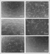
SEM images of collagen sheets seeded with different cell lines. (a) HUVECs and ASCs after six days of cultivation; (b) HUVECs after six days of cultivation; (c) HUVECs and ASCs after 19 days of cultivation; (d) HUVECs after 19 days of cultivation. Arrows: collagen fiber alignment. Circle: non-oriented collagen fibers.
Figure 4
Figure 4
AFM measurement of cryo-sections from 3D collagen sheets laden with a co-culture of ASCs and HUVECs after 19 days of incubation. (a) Young’s modulus distribution in a region of aligned collagen fibers; (b) bright-field optical microscopy image, showing the collagen sheet (grey background), the AFM cantilever (dark triangle), and the region where the 100 µm × 100 µm overview image displayed below (e) was recorded; (c) Young’s modulus distribution in a region of randomly oriented collagen fibers; (d) contact mode AFM image (height image) in the region of aligned collagen fibers; (e) 100 µm × 100 µm AFM overview image (cantilever deflection image); (f) AFM image in the region of randomly oriented collagen fibers (height image). White arrows: aligned collagen fibers, black arrow: random fiber orientation.
Figure 5
Figure 5
AFM measurement of cryo-sections from collagen sheets laden with HUVECs after 19 days of incubation. (a) Young’s modulus distribution in a region of aligned collagen fibers; (b) bright-field optical microscopy image, showing the collagen sheet (grey background), the AFM cantilever (dark triangle), and the region where the 100 µm × 100 µm overview image displayed below (e) was recorded; (c) Young’s modulus distribution in a region of randomly oriented collagen fibers; (d) contact mode AFM image (height image) in the region of aligned collagen fibers; (e) 100 µm × 100 µm AFM overview image (cantilever deflection image); (f) AFM image in the region of randomly oriented collagen fibers (height image). White arrow: aligned collagen fibers, black arrow: random fiber orientation.
Figure 6
Figure 6
Expression of selected angiogenic factors in cell-laden collagen sheets. Cell culture supernatants were collected after 6 and 19 days of cultivation and analyzed using the Proteome Profiler Human Angiogenesis Array Kit for potential growth factors. As a control, Endothelial Cell Growth Medium 2 (containing angiogenic growth factors) was used to validate the assay. The * shows the significant difference between the values. Therefore, the data were analyzed using one-way Analysis of Variance (ANOVA). Probability values (***) p < 0.001 were considered the most significant.
Figure 7
Figure 7
Bright-field microscopy images of the HUVECs tube formation assay after 4 h of incubation with supernatants obtained from the cultivation of different cell-laden 3D collagen sheets. (a) Treated with supernatant from 3D construct laden with HUVECs and ASCs after six days of cultivation; (b) Treated with supernatant from 3D HUVECs construct after six days of cultivation; (c) Treated with supernatant from 3D HUVECs and ASCs construct after 19 days of cultivation; (d) Treated with supernatant from 3D HUVECs construct after 19 days of cultivation; (e) positive control cultivated in Endothelial Cell Growth Medium 2 containing angiogenic growth factors; (f) negative control using AIM V cytokine and serum-free medium for incubation.
Figure 8
Figure 8
Quantitative analysis of the tube formation assay. (a) Cell-covered area per well; (b) total number of branching points per well; (c) total number of tubes per well. The cell culture supernatants were collected after different time points (days 6, 12, and 19) to investigate their angiogenic potential. Error bars indicate the standard deviation.

References

    1. Schreml S., Szeimies R.-M., Prantl L., Landthaler M., Babilas P. Wound healing in the 21st century. J. Am. Acad. Dermatol. 2010;63:866–881. doi: 10.1016/j.jaad.2009.10.048. - DOI - PubMed
    1. de Carvalho C.K.L., Fernandes B.L., de Souza M.A. Autologous matrix of platelet-rich fibrin in wound care settings: A systematic review of randomized clinical trials. J. Funct. Biomater. 2020;11:31. doi: 10.3390/jfb11020031. - DOI - PMC - PubMed
    1. Schultz G., Chin G., Moldawer L., Diegelmann R. Ch23 Mechanisms of Vascular Disease. University of Adelaide Press; Adelaide, Australia: 2011.
    1. Arnold F., West D.C. Angiogenesis in wound healing. Pharmacol. Ther. 1991;52:407–422. doi: 10.1016/0163-7258(91)90034-J. - DOI - PubMed
    1. Um J., Yu J., Dubon M.J., Park K.-S. Substance P and thiorphan synergically enhance angiogenesis in wound healing. Tissue Eng. Regen. Med. 2016;13:149–154. doi: 10.1007/s13770-016-9089-y. - DOI - PMC - PubMed