

PLGA Nanoparticles Uptake in Stem Cells from Human Exfoliated Deciduous Teeth and Oral Keratinocyte Stem Cells
- PMID: 35997447
- PMCID: PMC9397094
- DOI: 10.3390/jfb13030109
PLGA Nanoparticles Uptake in Stem Cells from Human Exfoliated Deciduous Teeth and Oral Keratinocyte Stem Cells
Abstract
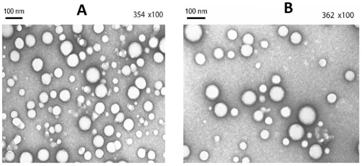
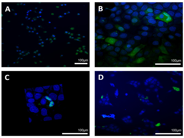
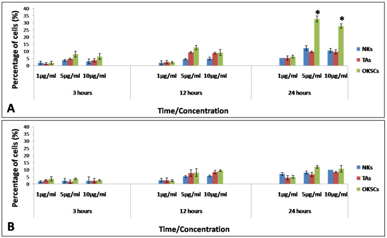
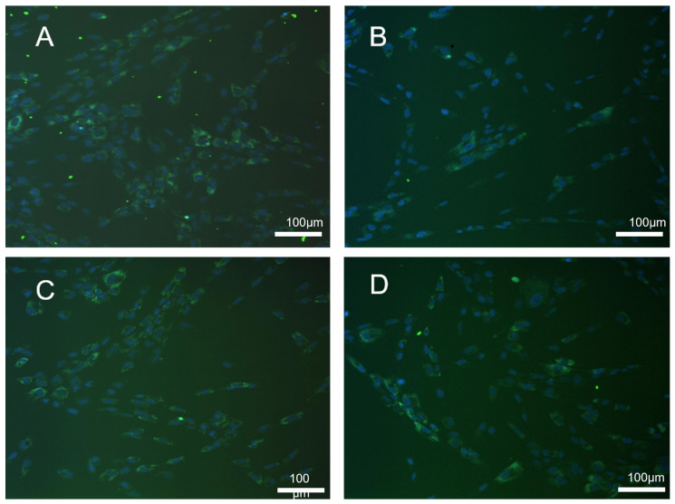
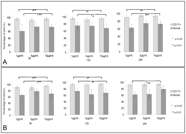
Polymeric nanoparticles have been introduced as a delivery vehicle for active compounds in a broad range of medical applications due to their biocompatibility, stability, controlled release of active compounds, and reduced toxicity. The oral route is the most used approach for delivery of biologics to the body. The homeostasis and function of oral cavity tissues are dependent on the activity of stem cells. The present work focuses, for the first time, on the interaction between two types of polymeric nanoparticles, poly (lactic-co-glycolic acid) or PLGA and PLGA/chitosan, and two stem cell populations, oral keratinocyte stem cells (OKSCs) and stem cells from human exfoliated deciduous teeth (SHEDs). The main results show that statistical significance was observed in OKSCs uptake when compared with normal keratinocytes and transit amplifying cells after 24 h of incubation with 5 and 10 µg/mL PLGA/chitosan. The CD117+ SHED subpopulation incorporated more PLGA/chitosan nanoparticles than nonseparated SHED. The uptake for PLGA/chitosan particles was better than for PLGA particles with longer incubation times, yielding better results in both cell types. The present results demonstrate that nanoparticle uptake depends on stem cell type, incubation time, particle concentration, and surface properties.
Keywords: PLGA nanoparticles; human exfoliated deciduous teeth; oral keratinocyte stem cells.
Conflict of interest statement
The authors declare no conflict of interest. The funders had no role in the design of the study; in the collection, analyses, or interpretation of data; in the writing of the manuscript; or in the decision to publish the results.
Figures
References
-
- Virlan M.J.R., Miricescu D., Totan A., Greabu M., Tanase C., Sabliov C.M., Constantin C., Calenic B. Current uses of poly (lactic-co-glycolic acid) in the dental field: A comprehensive review. J. Chem. 2015;2015:525832. doi: 10.1155/2015/525832. - DOI
-
- Calenic B., Ishkitiev N., Yaegaki K., Imai T., Costache M., Tovaru M., Parlatescu I. Characterization of oral keratinocyte stem cells and prospects of its differentiation to oral epithelial equivalents. Rom. J. Morphol. Embryol. 2010;51:641–645. - PubMed
Grants and funding
LinkOut - more resources
Full Text Sources
