. 2022 Sep;28(5):503-515.
doi: 10.5152/dir.2022.21339.

The changing landscape in epilepsy imaging: Unmasking subtle and unique entities

Affiliations

The changing landscape in epilepsy imaging: Unmasking subtle and unique entities

Mehmet E Adin et al. Diagn Interv Radiol. 2022 Sep.

Abstract

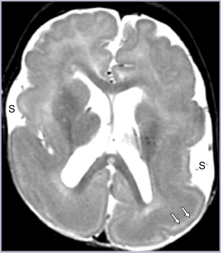
Dramatic changes have occurred recently in the field of epilepsy, including a fundamental shift in the etiology of epileptogenic substrates found at surgery. Hippocampal sclerosis is no longer the most common etiology found at epilepsy surgery and this decrease has been associated with an increase in the incidence of focal cortical dysplasia and encephaloceles. Significant advances have been made in molecular biology and genetics underlying the basis of malformations of cortical development, and our ability to detect epileptogenic abnormalities with MR imaging has markedly improved. This article begins with a discussion of these trends and reviews imaging techniques essential for detecting of subtle epilepsy findings. Representative examples of subtle imaging findings are presented, which are often overlooked but should not be missed. These include temporal lobe encephaloceles, malformations of cortical development (and especially focal cortical dysplasia), hippocampal sclerosis, hippocampal malformation (also known as HIMAL), ulegyria, autoimmune encephalitis, and Rasmussen's encephalitis. Recent findings on the pathophysiology and genetic underpinnings of several causes of localization-related epilepsy are incorporated. For instance, it has been recently found that focal cortical dysplasia IIb, tuberous sclerosis, hemimegalencephaly, and gangliogliomas are all the result of mutations of the mTOR pathway for cell growth.


Conflict of interest statement

Conflict of interest disclosure The authors declared no conflicts of interest.

Figures

Figure 1.
Figure 1.
(a) Subtle hippocampal sclerosis, with mild left hippocampal volume loss without associated hyperintense signal changes, is demonstrated on coronal T2W imaging at level of the hippocampal body. The alternating gray–white bands representing the hippocampal internal architecture are decreased on the left (arrow, compare with diagram in Figure 1c). (b) In a different patient, more typical changes of hippocampal sclerosis are present, with a small hyperintense hippocampus (arrow) associated with ipsilateral temporal lobe atrophy on a coronal T2W image. (c) Idealized coronal T2W diagram depicts many of the imaging features associated with hippocampal sclerosis, including hippocampal atrophy, hippocampal T2W signal abnormality, and loss of internal white matter architecture within the gray matter of the hippocampus (arrow). Associated temporal lobe atrophy is illustrated, associated with dysmyelination changes (arrowheads), as demonstrated by white matter hyperintensity and blurring of the gray–white matter junction.
Figure 2.
Figure 2.
Hippocampal malrotation (HIMAL). A 3-year-old boy with febrile and infantile seizures. Coronal T2W imaging (a) and diagram (b) demonstrate findings associated with HIMAL—a rounded hippocampal shape with a unilateral malrotated left hippocampus with blurred hippocampal internal architecture (black arrowheads), vertical collateral sulcus (long white arrow; compared to normal contralateral side, white arrowhead), and an abnormal temporal horn configuration (short white arrow).
Figure 3.
Figure 3.
Temporal lobe encephalocele. A 36-year-old female with intractable complex partial seizures since age 28. The left temporal pole encephalocele (arrows) is easy to overlook without using a systematic approach to interpretations (coronal T2W and FLAIR Imaging, (a) and (b), respectively). The patient underwent left temporal pole resection, which confirmed an encephalocele, with pathologic changes of reactive gliosis. The patient has been seizure-free since resection.
Figure 4.
Figure 4.
Autoimmune-mediated encephalitis (AME). A 57-year-old female presented with progressive confusion over 3 days, which evolved to visual hallucinations and then complex partial seizures. On coronal images, hippocampi are slightly enlarged and hyperintense bilaterally (arrows) on T2W imaging (a), consistent with limbic encephalitis that was due to voltage-gated potassium channel complex antibodies. Despite immunotherapy and steroids, the patient was left with memory loss and intractable epilepsy. (b). AME classification schemes, using a diagram of a neuron with cell membrane in black and intracellular compartment green. AME can be categorized by (1) cellular location of antigens—membranous versus intracellular, (2) site of brain encephalitis—limbic versus rarely limbic, and (3) association with neoplastic disease—paraneoplastic versus rarely paraneoplastic.
Figure 5.
Figure 5.
Hemimegalencephaly, partial. Axial T1W image displays an enlarged posterior left hemisphere, with dysplastic hyperintense cortex associated with blurred gray–white junction (white arrows) and periventricular heterotopia (black arrowheads), consistent with partial hemimegalencephaly. Identification is more challenging when hemimegalencephaly involves only a portion of a hemisphere instead of the entire hemisphere.
Figure 6.
Figure 6.
Focal cortical dysplasia. A 28-year-old female with medically refractory seizures since age 4, consisting of left arm tingling and dystonia; EEG suggested midline central origin. MRI was initially interpreted as normal, but reevaluation after knowledge of semiology and EEG findings revealed minimally thickened, hyperintense gyrus with blurred gray–white interface, in the right paracentral lobule (white arrow) on axial T1W (a) and axial FLAIR (b) images. Very subtle radial band (arrowhead) is present on axial FLAIR (c). This was confirmed on an oblique coronal reformatted image (d), showing subtle subcortical hyperintensity (arrow) with a radial band to the ventricle (arrowheads). F-18 FDG-PET also confirmed this abnormal region. (e). Focal cortical dysplasia type IIb in a different patient with seizure onset at age 15 consisted of episodes of flashing lights. A right parietal lesion was resected and showed both dysmorphic neurons (black arrow) and balloon cells with eccentric nucleus (arrowhead) on photomicrograph (original magnification, 20×; hematoxylin-eosin (H-E) stain). F-18 FDG-PET, F-18-fluorodeoxyglucose positron emission tomography.
Figure 7.
Figure 7.
Focal cortical dysplasia (FCD). A 2-year-old boy with intractable right arm and leg clonic seizures with FCD IIa. Axial T1W image (a) demonstrates markedly disorganized perirolandic sulcal pattern (compared to normal right central sulcus), pronounced thickening of the cortex with hyperintensity, and blurring of the cortical-white matter junction (arrows). Intraoperative photograph of brain surface with subdural grid electrodes (b) found marked distortion of brain motor representation, compared to the typical motor homunculus pattern (motor activity represented by electrodes in green; seizure activity represented by electrodes in blue, which were located adjacent to the interhemispheric fissure). Seizure freedom was obtained after functional hemispherectomy.
Figure 8.
Figure 8.
Tuberous sclerosis complex. (a) Hyperintense cortical tubers in the subcortical white matter with associated with radial bands (arrows) on coronal FLAIR. (b, c) Different patient with tuberous sclerosis. (b) Subependymal periventricular nodule (note signal void representing calcification on axial SWI [susceptibility weighted image]). (c) Enhancing (left greater than right) subependymal giant cell astrocytomas (arrow points to left SEGA on axial postcontrast T1W MP-RAGE [magnetization-prepared rapid acquisitive gradient echo] image). SEGA, subependymal giant cell astrocytoma.
Figure 9.
Figure 9.
Lissencephaly. There is generalized smooth thickened cortex, with open Sylvian fissures (S) with poor gray–white junction and paucity of white matter on axial T2W imaging, representing classic lissencephaly. Note the hyperintense sparse cell zone with increased water content on T2W imaging (white arrows), a feature of lissencephaly which can sometimes be misinterpreted as indicating double cortex syndrome (band heterotopia).
Figure 10.
Figure 10.
Polymicrogyria. Right polymicrogyria is nicely depicted on sagittal (a) and coronal (b) T1W imaging as numerous small gyri (arrows). The perisylvian location, as seen here, is the most common region affected and often results in fusion of the Sylvian fissure with the central sulcus (arrowheads), as in this case. Findings may be unilateral or bilateral; when bilateral asymmetric, it may be easy to overlook a subtle contralateral abnormality (as in this case, with very subtle thickening and increased nodularity of the contralateral left insula and frontal operculum).
Figure 11.
Figure 11.
Hypothalamic hamartoma. An 8-year-old boy with complex partial seizure. Coronal T1W image demonstrates a hypothalamic mass (arrow) encompassing the mammillary body that was isointense to gray matter on multiple sequences, consistent with a hypothalamic hamartoma. A patterned interpretation approach must include the evaluation of the hypothalamus and mamillary body in patients with epilepsy to detect subtle lesions.
Figure 12.
Figure 12.
Ulegyria. A 11-year-old boy with family history of coagulation disorder had infarcts at 2 weeks of age and perinatal seizures. Coronal FLAIR (a) and T2W imaging (b) demonstrate typical bilateral watershed-territory ulegyria. Note the atrophic gyri with mushroom-like configuration at the dome (black arrow), dilatation of CSF at the depth of the sulci (white arrow), subcortical white matter signal changes, and bilateral parasagittal locations. CSF, cerebrospinal fluid.
Figure 13.
Figure 13.
Rasmussen’s encephalitis. A 5-year-old girl with 1-year history of right body twitching that developed into right body motor partial status epilepticus. Left parietal white matter hyperintensity (black arrow) was present on initial axial FLAIR MRI (a), which extended into adjacent white and gray matter as subtle hyperintensity, with loss of gray–white matter junction (white arrows), 1 year later (b). MR imaging findings were compatible with Rasmussen’s progression and ictal changes, with possible concomitant FCD. F-18 FDG PET-CT (not shown) revealed hypometabolism in the left cerebral hemisphere. Biopsy revealed perivascular distribution of CD3/8 positive T-cells and microglial cells (CD68) with type Ib FCD, consistent with Rasmussen encephalitis in the setting of dual pathology. MRI, magnetic resonance imaging; F-18 FDG-PET, F-18-fluorodeoxyglucose positron emission tomography; FCD, focal cortical dysplasia.

Similar articles

Cited by

References

    1. Barkovich AJ, Dobyns WB, Guerrini R. Malformations of cortical development and epilepsy. Cold Spring Harb Perspect Med. 2015;5(5):a022392. 10.1101/cshperspect.a022392) - DOI - PMC - PubMed
    1. Muhlhofer W, Tan YL, Mueller SG, Knowlton R. MRI-negative temporal lobe epilepsy-what do we know? Epilepsia. 2017;58(5):727 742. 10.1111/epi.13699) - DOI - PubMed
    1. Jehi L, Friedman D, Carlson C.et al. The evolution of epilepsy surgery between 1991 and 2011 in nine major epilepsy centers across the United States, Germany, and Australia. Epilepsia. 2015;56(10):1526 1533. 10.1111/epi.13116) - DOI - PMC - PubMed
    1. Richardson MP. Large scale brain models of epilepsy: dynamics meets connectomics. J Neurol Neurosurg Psychiatry. 2012;83(12):1238 1248. 10.1136/jnnp-2011-301944) - DOI - PubMed
    1. Spencer SS. Neural networks in human epilepsy: evidence of and implications for treatment. Epilepsia. 2002;43(3):219 227. 10.1046/j.1528-1157.2002.26901.x) - DOI - PubMed

MeSH terms

Substances