Review
. 2022 Aug;57(4):399-411.
doi: 10.1080/10409238.2022.2113030. Epub 2022 Aug 23.

What makes functional amyloids work?

Affiliations
Review

What makes functional amyloids work?

Ansgar B Siemer. Crit Rev Biochem Mol Biol. 2022 Aug.

Abstract

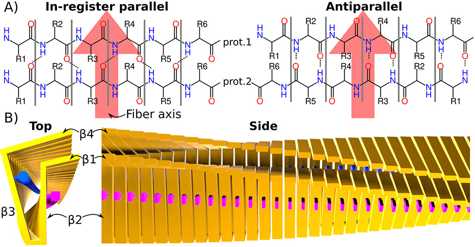
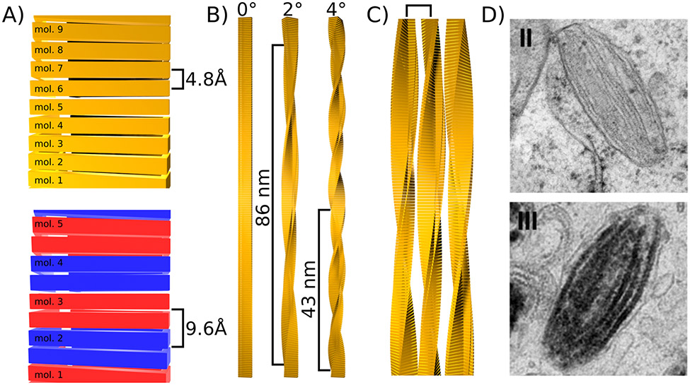
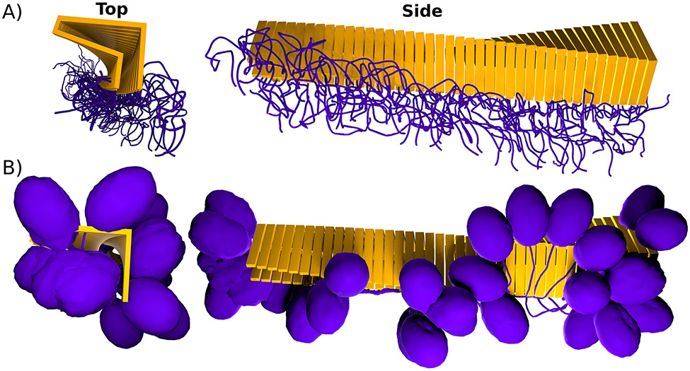
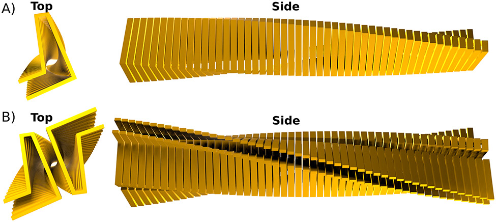
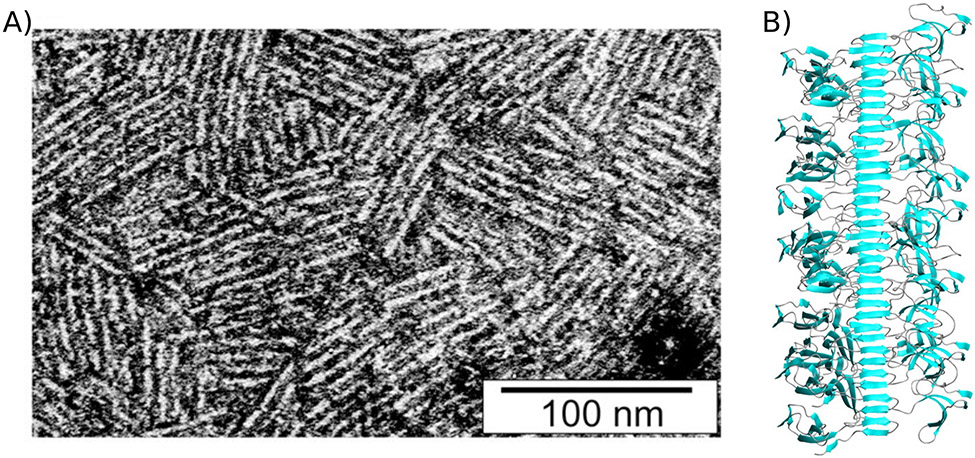
Although first described in the context of disease, cross-β (amyloid) fibrils have also been found as functional entities in all kingdoms of life. However, what are the specific properties of the cross-β fibril motif that convey biological function, make them especially suited for their particular purpose, and distinguish them from other fibrils found in biology? This review approaches these questions by arguing that cross-β fibrils are highly periodic, stable, and self-templating structures whose formation is accompanied by substantial conformational change that leads to a multimerization of their core and framing sequences. A discussion of each of these properties is followed by selected examples of functional cross-β fibrils that show how function is usually achieved by leveraging many of these properties.

Keywords: Functional amyloid; cross-β motif; protein aggregation; protein fibrils; structure–function relationship.


Figures

Figure 1:
Figure 1:. Core structure of cross-β fibrils.
A) Schematics of in-register cross-β (found in most cross-β fibrils) and antiparallel cross-β structures. Two layers formed by two monomers are shown. The fibril axis is indicated with a large red arrow. In the parallel-in register motif, the same residues from each monomer are right on top of each other, which is not the case in the antiparallel (or out of register parallel) case. B) Idealized in-register, parallel cross-β fibril. The protein backbone of each monomer is represented by yellow bars. Two examples of side chains are illustrated as blue circles and pink squares. 4 β-strands connected via kinks and turns can be identified in the top view. The side view shows the large continuous β-sheets formed by strands β2 and β4 from each monomer.
Figure 2:
Figure 2:. Levels of periodicity in cross-β fibrils.
A) Periodicity along the fibril axis caused by the repetition of monomers. In the simplest case when every strand of a β-sheet is formed by a different monomer, the periodicity is 4.8 Å (top). Otherwise the this periodicity is multiple of 4.8 Å. For example when one monomer provides two strands of the same β-sheet as illustrated with alternating blue and red monomers (bottom). B) Periodicity along the fibril axis caused by a slight rotation of each monomer around the fibril axis (yaw). Three examples with rotation angles of 0°, 2°, and 4° are given and the resulting periodicity is indicated. C) Periodicity induced by lateral association of fibrils. A square bracket indicates the resulting periodicity. D) Example of lateral association of Pmel17 fibrils during stage II of melanosome formation (top), to which melanin binds during stage III (bottom). Images from Hurbain and co-workers (2008) Copyright 2008 National Academy of Sciences.
Figure 3:
Figure 3:. Cross-β formation causes multimerization of core and framing sequences.
A) Idealized cross-β fibril with intrinsically disordered framing sequences (purple) that form an entropic brush at the surface of the fibril. B) Idealized cross-β fibril with globular domain (purple) connected to the cross-β core (yellow) via a linker.
Figure 4:
Figure 4:. Cross-β fibrils can be made of one or multiple protofilaments.
A) View down the fibril axis (Top) and along the fibril axis (Side) of an idealized cross-β core formed by a single (proto)filament. In such a fibril there is only one monomer per layer. B) Idealized cross-β fibril that is made of two protofilaments. The top view shows nicely how this fibil has two identical monomers per layer.
Figure 5:
Figure 5:. Class I hydrophobin rodlet structures are stabilized by a cross-β core.
A) transmission electron micrograph of negatively stained EASΔ15 rodlet monolayers. Image from Morris et. al. 2011 under a CC BY 4.0 license (https://creativecommons.org/licenses/by/4.0/). B) Model of EASΔ15 fibrils showing the fibril core and globular, amphipathic framing sequences. Image from Macindoe et al. 2012.

Similar articles

Cited by

References

    1. Alberti S, Hyman AA. 2021. Biomolecular condensates at the nexus of cellular stress, protein aggregation disease and ageing. Nat Rev Mol Cell Biol. 22(3):196–213. doi:10.1038/s41580-020-00326-6. - DOI - PubMed
    1. Alberts B, Johnson A, Lewis J, Raff M, Roberts K, Walter P. 2002. The Self-Assembly and Dynamic Structure of Cytoskeletal Filaments. Mol Biol Cell 4th Ed. https://www.ncbi.nlm.nih.gov/books/NBK26862/.
    1. Andersson M, Johansson J, Rising A. 2016. Silk Spinning in Silkworms and Spiders. Int J Mol Sci. 17(8):1290. doi:10.3390/ijms17081290. - DOI - PMC - PubMed
    1. Anfinsen CB. 1973. Principles that govern the folding of protein chains. Science. 181(4096):223–230. doi:10.1126/science.181.4096.223. - DOI - PubMed
    1. Baba M, Nakajo S, Tu P-H, Tomita T, Nakaya K, Lee VM-Y, Trojanowski JQ, Iwatsubo T. 1998. Aggregation of α-synuclein in Lewy bodies of sporadic Parkinson’s disease and dementia with Lewy bodies. Am J Pathol. 152(4):879–884. - PMC - PubMed

Publication types

LinkOut - more resources