. 2022 Aug 23;117(1):40.
doi: 10.1007/s00395-022-00948-1.

Mitochondrial calpain-1 activates NLRP3 inflammasome by cleaving ATP5A1 and inducing mitochondrial ROS in CVB3-induced myocarditis

Affiliations

Mitochondrial calpain-1 activates NLRP3 inflammasome by cleaving ATP5A1 and inducing mitochondrial ROS in CVB3-induced myocarditis

Xiaoxiao Liu et al. Basic Res Cardiol. .

Abstract

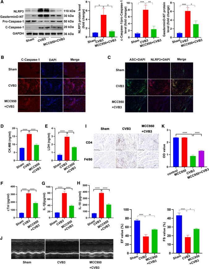
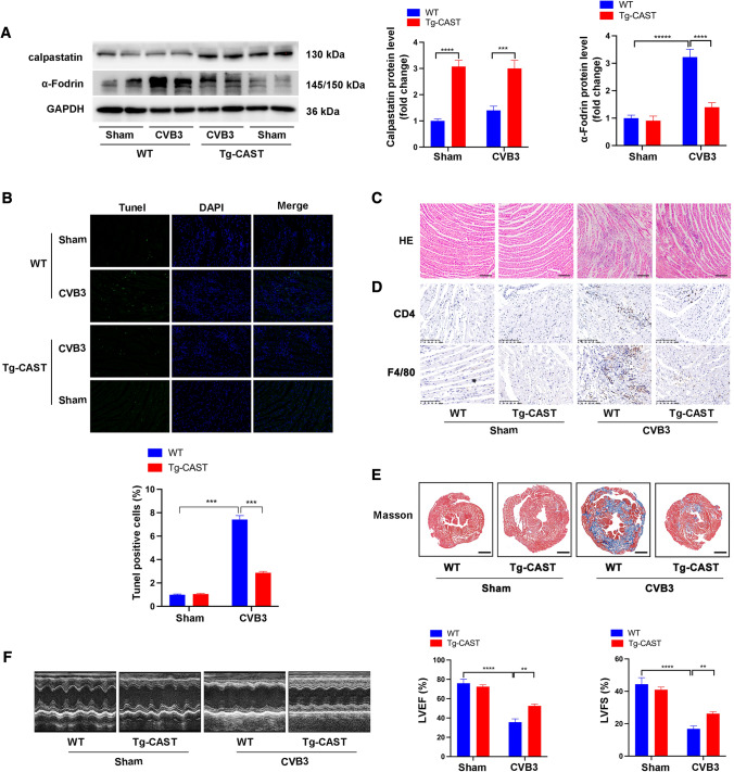
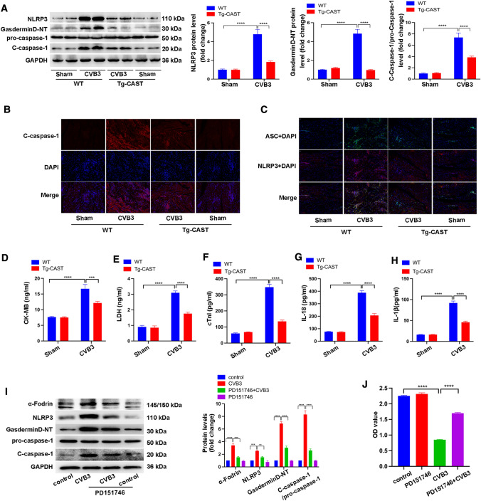
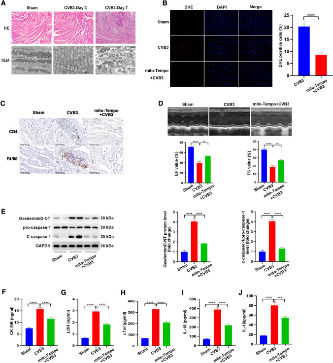
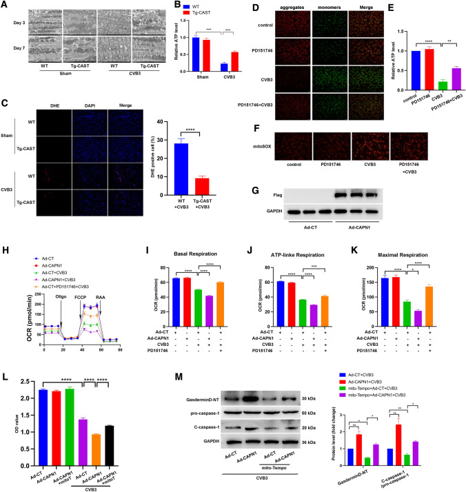
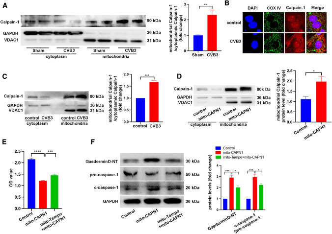
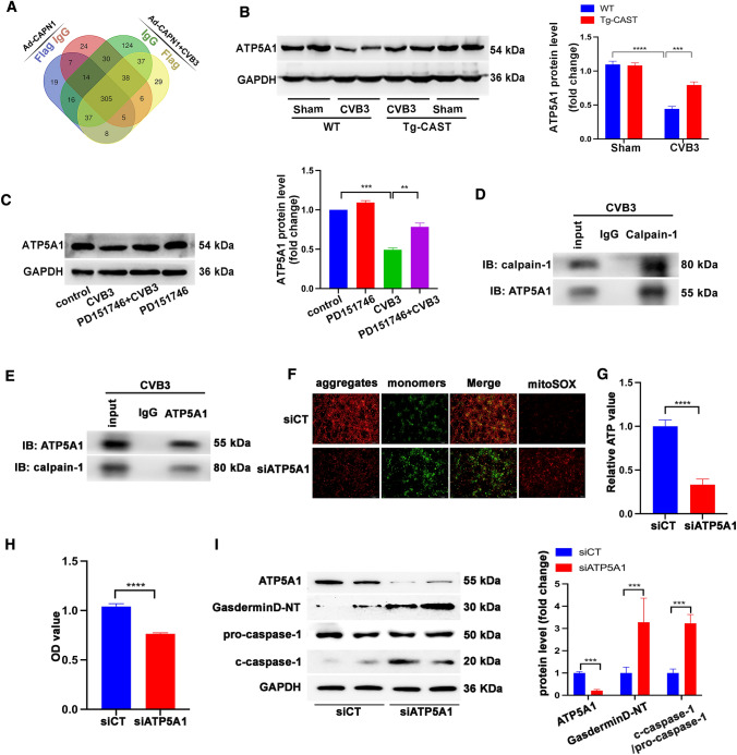
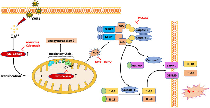
Treatment options for myocarditis are currently limited. Inhibition of calpains has been shown to prevent Coxsackievirus B3 (CVB3)-induced cardiac injuries, but the underlying mechanism of action of calpains has not been elucidated. We investigated whether NOD-, LRR-, and pyrin domain-containing 3 (NLRP3) inflammasome participated in CVB3-induced myocarditis, and investigated the effects of calpain-1 on CVB3-induced cardiac injury. NLRP3 inflammasome was activated in CVB3-infected hearts, evidenced by elevated protein levels of NLRP3, N-terminal domain of Gasdermin D, and cleaved caspase-1, and the increased co-localization of NLRP3 and apoptosis-associated speck-like protein. The intraperitoneal administration of MCC950, a selective inhibitor of the NLRP3 inflammasome, led to decreased levels of serum creatine kinase-MB, cardiac troponin I, lactate dehydrogenase, interleukin-18, interleukin-1β, prevention of the infiltration of inflammatory cells, and improvement of cardiac function under CVB3 infection. Transgenic mice overexpressing the endogenous calpain inhibitor calpastatin (Tg-CAST mice) exhibited not only decreased apoptosis, inflammation, fibrosis, and enhanced cardiac function but also inhibition of NLRP3 inflammasome and pyroptosis. The selective inhibition of calpain-1 using PD151746 protected cardiomyocytes in vitro from CVB3 infection by downregulating NLRP3 inflammasome and, thus, preserved cell viability. Mechanistically, we showed that mitochondrial dysfunction preceded inflammatory response after CVB3 treatment and elimination of mitochondrial reactive oxygen species (ROS) using mitochondria-targeted antioxidants (mito-TEMPO) recapitalized the phenotype observed in Tg-CAST mice. Furthermore, the promotion or inhibition of calpain-1 activation in vitro regulated the mitochondrial respiration chain. Mito-TEMPO reversed calpain-1-mediated NLRP3 inflammation activation and cell death. We also found that mitochondrial calpain-1, which was increased after CVB3 stimulation, activated the NLRP3 inflammasome and resulted in cell death. Furthermore, ATP synthase-α (ATP5A1) was revealed to be the cleaving target of calpain-1 after CVB3 treatment. Downregulating ATP5A1 using ATP5A1-small interfering RNA impaired mitochondrial function, decreased cell viability, and induced NLRP3 inflammasome activation. In conclusion, CVB3 infection induced calpain-1 accumulation in mitochondria, and led to subsequent ATP5A1 cleavage, mitochondrial ROS overproduction, and impaired mitochondrial function, eventually causing NLRP3 inflammasome activation and inducing pyroptosis. Therefore, our findings established the role of calpain in viral myocarditis and unveiled its underlying mechanism of its action. Calpain appears as a promising target for the treatment of viral myocarditis.

Keywords: ATP5A1; Calpain; Mitochondrial ROS; NLRP3 inflammasome; Viral myocarditis.


Conflict of interest statement

The authors declared no conflicts of interest, financial, or otherwise.

Figures

Fig. 1
Fig. 1
Mouse models of myocarditis were established by intraperitoneally injecting mice with CVB3. To inhibit NLRP3 inflammasome activation in CVB3-infected mice, MCC950 was administrated intraperitoneally at a dose of 10 mg/kg daily starting at the 2 days before CVB3 treatment. Assays were carried out at 7 days after CVB3 infection. A Representative image of western blotting results and the quantitative analysis of NLRP3, Gasdermin D-NT, pro-caspase 1, c-caspase 1 in heart tissues. B, C Representative images of immunofluorescence staining showing the expression of c-caspase-1, NLRP3, and ASC in heart sections. D, E, F, G, H Levels of serum CK-MB, LDH, cTnI, IL-18, and IL-1β in mice subject to the indicated treatments. I Representative images of immunohistochemistry staining showing the cardiac infiltration of CD4-positive T lymphocytes and F4/80-positive macrophages. J Echocardiography images and quantitative statistics of ejection fraction (EF) and fractional shortening (FS). N = 6 mice in each group. K NRCMs were treated with MCC950 or CVB3 infection or both MCC950 and CVB3. After 48 h, CCK8 was used to assess cell viability in each group. N = 16 replicates from three independent experiments
Fig. 2
Fig. 2
CVB3-induced myocarditis models were constructed in calpastatin overexpression mice (Tg-CAST) and their wild-type littermates (WT). A Representative image of western blotting results and the quantitative analysis of calpastatin and α-Fodrin after CVB3 infection for 7 days. B Representative images and quantification of TUNEL-positive cells in heart section after CVB3 infection for 7 days. Data were collected from 18 fields from six mice in each group. C HE staining of heart sections showing local changes in different mice after CVB3 infection for 7 days; bar = 100 µm. D Representative images of immunohistochemistry staining showing cardiac infiltration of CD4 positive T lymphocytes and F4/80-positive macrophages after CVB3 infection for 7 days; bar = 100 µm. E Masson’s trichrome staining of heart sections showing fibrosis after CVB3 infection for 7 days.; bar = 500 mm. F Echocardiography images and quantitative statistics of EF and FS. N = 6 mice in each group
Fig. 3
Fig. 3
NLRP3 inflammasome activation and pyroptosis in Tg-CAST and WT mice with or without CVB3 infection. A Representative image of western blotting results and the quantitative analysis of NLRP3, gasdermin D-NT, pro-caspase 1, and c-caspase 1 in heart tissues. B, C Representative images of immunofluorescence staining showing the expression of c-caspase-1, NLRP3, and ASC in each group. D, E, F, G, H Serum levels of CK-MB, LDH, cTnI, IL-18, and IL-1β. N = 6 mice in each group. NRCMs were treated with PD151746 or CVB3 or both PD151746 and CVB3 for 48 h. I Representative image of western blotting results and the quantitative analysis of α-Fodrin, NLRP3, gasderminD-NT, pro-caspase-1, and c-caspase-1. N = 3 independent experiments. G CCK8 showing the viability of NRCMs after the indicated treatments. N = 24 from three independent experiments
Fig. 4
Fig. 4
To determine whether mitochondrial ROS participated in CVB3-induced myocarditis, mito-TEMPO at a dose of 0.7 mg/kg daily was administrated intraperitoneally starting at the 2 days before CVB3 treatment. Assays were conducted after CVB3 infection. A Representative images of HE staining and transmission electron microscopy showing changes in the histology and mitochondria of the heart on 3rd day or 7th day after CVB3 treatment. B DHE staining and quantification of positive DHE in nuclei. N = 4 mice from each group. C Representative images of immunohistochemistry staining showing cardiac infiltration of CD4-positive T lymphocytes and F4/80-positive macrophages. D Echocardiography images and quantitative analysis of EF and FS. E Representative image of western blotting results and quantitative analysis of gasdermin D-NT, pro-caspase-1, and c-caspase-1. F, G, H, I, J Serum levels of CK-MB, LDH, cTnI, IL-18, and IL-1β in mice subjected to different treatments. N = 6 mice in each group
Fig. 5
Fig. 5
A Transmission electron microscopy images showing the structure of cardiac mitochondria subjected to different treatments. B Relative ATP levels in the heart of WT or Tg-CAST mice with or without CVB3 infection for 7 days. N = 6 mice in each group. C Representative images of DHE staining. After oxidization, DHE intercalates into DNA and stains the nuclei red. The percentages of DHE-positive cells were quantified. N = 4 mice. D Representative images of JC-1 staining of NRCMs showing the mitochondrial membrane potential in each group. N = 3 independent experiments. E Relative ATP levels in NRCMs after treatment with CVB3 or PD151746. N = 3 independent experiments. F Representative images of mitochondrial ROS in NRCMs stained by mitoSOX indicator. N = 3 independent experiments. G NRCMs were infected with CAPN1-expressing adenovirus (Ad-CAPN1) or control adenovirus (Ad-CT) at an MOI (multiplicity of infection) of 50 for 48 h, and the Flag-tag was then detected by western blotting to show the effective infection of Ad-CAPN1. N = 3 independent experiments. H NRCMs were infected with Ad-CT or Ad-CAPN1 for 48 h with or without CVB3 treatment. PD151746 was added to some cells to inhibit the activation of calpain-1. After treatment, oxygen consumption rate (OCR) was detected using the Seahorse platform. I Basal respiration; J ATP-linked respiration; K maximal respiration. N = 17 replicates in each group. L NRCMs were infected with Ad-CT or Ad-CAPN1 for 48 h with or without CVB3 treatment. Mito-TEMPO was added to NRCMs treated with Ad-CAPN1 and CVB3. CCK8 shows cell viability. N = 16 replicates from three independent experiments. M Representative image of western blotting bands and the quantitative analysis of gasdermin D-NT, pro-caspase-1, and c-caspase-1 under different treatments. N = 3 independent experiments
Fig. 6
Fig. 6
A Mitochondria was isolated from cytoplasm in CVB3-infected hearts or control hearts, and then, protein levels of calpain-1 were detected in each fraction. N = 6 mice in each group. B Immunofluorescence staining showing the co-localization of calpain-1 and mitochondria in NRCMs infected with CVB3. C calpain-1 levels in mitochondria isolated from CVB3-infected NRCMs. N = 3 independent experiments. D Representative image of western blotting results and the quantitative analysis of calpain-1 in cytoplasm or mitochondria in AC16 cells transfected with a plasmid expressing mitochondria-targeted calpain-1 (mito-CAPN1) or control plasmid. N = 3 independent experiments. E Viability of AC16 cells transfected with mito-CAPN1 and incubated with mito-TEMPO as determined by the CCK8 assay. N = 24 replicates from three independent experiments. F Representative image of western blotting results and the quantitative analysis of gasdermin D-NT, pro-caspase-1, and c-caspase-1. N = 3 independent experiments
Fig. 7
Fig. 7
A Venn diagram showing eight proteins coexisting in NRCMs treated with Ad-CAPN1 or co-treated with Ad-CAPN1 and CVB3, as determined by co-immunoprecipitation using anti-IgG or anti-Flag antibody and subjected to mass spectrometry to determine calpain-1-interacting proteins. B Representative image of western blotting results and the quantitative analysis of ATP synthase-α (ATP5A1) in mice. N = 6 mice in each group. C Representative image of western blotting results and the quantitative analysis of ATP synthase-α (ATP5A1) in NRCMs subjected to the indicated treatments. N = 3 independent experiments. D Immunoprecipitation assay using anti-calpain-1 antibody or anti-IgG antibody (negative control antibody). Calpain-1 and ATP5A1 levels in co-immunoprecipitated samples. E Immunoprecipitation assay using anti-ATP5A1 antibody or anti-IgG antibody (negative control antibody). Calpain-1 and ATP5A1 levels in co-immunoprecipitated samples. F NRCMs were transfected with siRNA-Scramble (siCT) or siRNA-ATP5A1 (siATP5A1), and then, JC-1 staining indicated the mitochondrial membrane potential and mitoSOX indicated mitochondrial ROS. G ATP levels in NRCMs transfected with siCT or siATP5A1. N = 6 replicates from three independent experiments. H CCK8 assay showing the viability of NRCMs. N = 16 replicates from three independent experiments. I Representative image of western blotting results and the quantitative analysis of ATP5A1, gasdermin D-NT, pro-caspase-1, and c-caspase-1 in siCT- or siATP5A1-infected NRCMs. N = 3 independent experiments
Fig. 8
Fig. 8
Calpain-1 activated under CVB3 stimulation promoted NLRP3 inflammasome-dependent pyroptosis through ATP5A1 cleavage and ROS overproduction in mitochondria

References

    1. Abbate A, Kontos MC, Grizzard JD, Biondi-Zoccai GG, Van Tassell BW, Robati R, Roach LM, Arena RA, Roberts CS, Varma A, Gelwix CC, Salloum FN, Hastillo A, Dinarello CA, Vetrovec GW, Investigators V-A. Interleukin-1 blockade with anakinra to prevent adverse cardiac remodeling after acute myocardial infarction (Virginia Commonwealth University Anakinra Remodeling Trial [VCU-ART] Pilot study) Am J Cardiol. 2010;105(1371–1377):e1371. doi: 10.1016/j.amjcard.2009.12.059. - DOI - PubMed
    1. Abbate A, Toldo S, Marchetti C, Kron J, Van Tassell BW, Dinarello CA. Interleukin-1 and the Inflammasome as therapeutic targets in cardiovascular disease. Circ Res. 2020;126:1260–1280. doi: 10.1161/CIRCRESAHA.120.315937. - DOI - PMC - PubMed
    1. Cavalli G, Pappalardo F, Mangieri A, Dinarello CA, Dagna L, Tresoldi M. Treating life-threatening myocarditis by blocking Interleukin-1. Crit Care Med. 2016;44:e751–754. doi: 10.1097/CCM.0000000000001654. - DOI - PubMed
    1. Chen B, Zhao Q, Ni R, Tang F, Shan L, Cepinskas I, Cepinskas G, Wang W, Schiller PW, Peng T. Inhibition of calpain reduces oxidative stress and attenuates endothelial dysfunction in diabetes. Cardiovasc Diabetol. 2014;13:88. doi: 10.1186/1475-2840-13-88. - DOI - PMC - PubMed
    1. Chow LH, Gauntt CJ, McManus BM. Differential effects of myocarditic variants of Coxsackievirus B3 in inbred mice. A pathologic characterization of heart tissue damage. Lab Invest. 1991;64:55–64. - PubMed

Publication types

MeSH terms