Randomized Controlled Trial
. 2022 Oct 10;7(19):e158314.
doi: 10.1172/jci.insight.158314.

A randomized placebo-controlled trial of nicotinamide riboside and pterostilbene supplementation in experimental muscle injury in elderly individuals

Affiliations
Randomized Controlled Trial

A randomized placebo-controlled trial of nicotinamide riboside and pterostilbene supplementation in experimental muscle injury in elderly individuals

Jonas Brorson Jensen et al. JCI Insight. .

Abstract

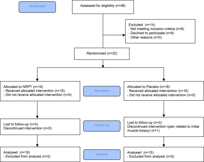
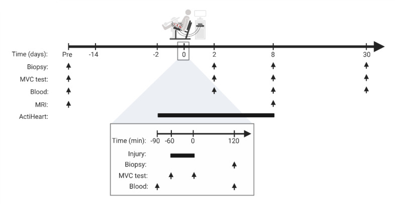
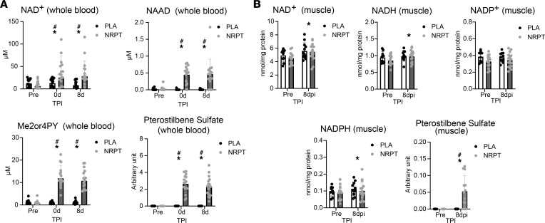
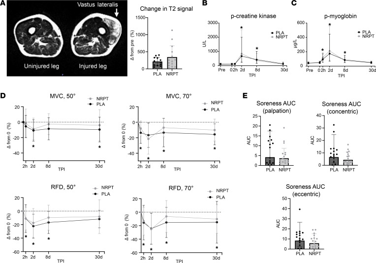
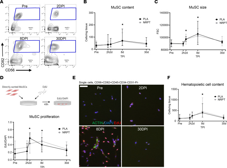
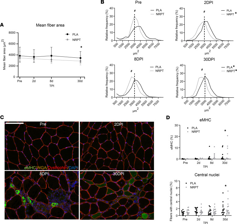
BACKGROUNDDuring aging, there is a functional decline in the pool of muscle stem cells (MuSCs) that influences the functional and regenerative capacity of skeletal muscle. Preclinical evidence has suggested that nicotinamide riboside (NR) and pterostilbene (PT) can improve muscle regeneration, e.g., by increasing MuSC function. The objective of this study was to investigate if supplementation with NR and PT (NRPT) promotes skeletal muscle regeneration after muscle injury in elderly individuals by improved recruitment of MuSCs.METHODSThirty-two elderly individuals (55-80 years of age) were randomized to daily supplementation with either NRPT (1,000 mg NR and 200 mg PT) or matched placebo. Two weeks after initiation of supplementation, skeletal muscle injury was induced by electrically induced eccentric muscle work. Skeletal muscle biopsies were obtained before, 2 hours after, and 2, 8, and 30 days after injury.RESULTSA substantial skeletal muscle injury was induced by the protocol and associated with release of myoglobin and creatine kinase, muscle soreness, tissue edema, and a decrease in muscle strength. MuSC content, proliferation, and cell size revealed a large demand for recruitment after injury, but this was not affected by NRPT. Furthermore, histological analyses of muscle fiber area, central nuclei, and embryonic myosin heavy chain showed no NRPT supplementation effect.CONCLUSIONDaily supplementation with 1,000 mg NR and 200 mg PT is safe but does not improve recruitment of the MuSC pool or other measures of muscle recovery in response to injury or subsequent regeneration in elderly individuals.TRIAL REGISTRATIONClinicalTrials.gov NCT03754842.FUNDINGNovo Nordisk Foundation (NNF17OC0027242) and Novo Nordisk Foundation CBMR.

Keywords: Aging; Human stem cells; Muscle Biology; Skeletal muscle.


Figures

Figure 1
Figure 1. CONSORT flow chart.
Figure 2
Figure 2. Study overview.
Participants were examined before initiation of NRPT or matched placebo supplementation (Pre). Supplementation started 14 days before induction of skeletal muscle injury and continued until 30 days after injury. Skeletal muscle injury was induced at time point 0 by electrically induced contractions combined with eccentric work in a dynamometer (200 repetitions, 100 slow and 100 fast). The bottom line gives a detailed description of the injury day. The arrows indicate timing of skeletal muscle biopsy, MVC test, blood samples, and MR imaging, respectively. Bars indicate time span. Skeletal muscle biopsies were obtained from the uninjured leg prior to starting supplementation and from the injured leg after induction of injury. MVC, maximal voluntary contraction.
Figure 3
Figure 3. NAD+ metabolites and pterostilbene sulfate in whole blood and skeletal muscle.
(A) Analysis of whole blood revealed an increase in NAD+, nicotinic acid adenine dinucleotide (NAAD), N1-methyl-2-pyridone-5-carboxamide/N1-Methyl-4-pyridone-3-carboxamide (Me2/4PY), and pterostilbene sulfate following NRPT supplementation. (B) NAD+, NADH, and NADPH were increased in skeletal muscle after injury, whereas NADP+ was not. NRPT supplementation was able to increase pterostilbene sulfate in skeletal muscle tissue. Data are expressed as mean ± SD and were compared using repeated-measurement mixed-model analysis. For pterostilbene sulfate, each group was tested separately using Friedman’s test, and for supplementation effect, groups were compared at each time point after injury with the Mann-Whitney test. Whole blood: NRPT, n = 16; PLA, n = 15. Muscle NAD+ metabolites: NRPT, n = 16; PLA, n = 14. Muscle pterostilbene sulfate: NRPT, n = 15; PLA, n = 11. #P < 0.05 between groups; *P < 0.05 vs. Pre. TPI, time after injury.
Figure 4
Figure 4. Clinical parameters associated with skeletal muscle injury.
(A) T2-weighted axial image of the thigh shows increased signal intensity in the vastus lateralis part of the quadriceps femoris muscle 8 days after injury, with no difference between groups (NRPT, n = 14; PLA, n = 12). (B) P-creatinine kinase, (C) p-myoglobin, (D) maximal voluntary contraction and rate of force development at 50- and 70-angle degrees (MVC, 50°/70° and RFD, 50°/70°), and (E) muscle soreness did response to injury but not to NRPT supplementation (NRPT, n = 16; PLA, n = 15). Data are expressed as geometric mean ± SD, with the exception of MVC and RFD data in D, which are expressed as mean ± SD. Data were compared using repeated-measurement mixed-model analysis, with the exception of MRI and muscle soreness data, which were compared using a Student’s t test. *P < 0.05 vs. Pre. TPI, time after injury; MVC, maximal voluntary contraction; RFD, rate of force development; AUC, area under the curve.
Figure 5
Figure 5. Muscle stem cell response to skeletal muscle injury.
(A) CD56 (neural cell adhesion molecule 1) versus CD82 (KAI1) contour flow plots of single CD45CD31CD34PI cells from skeletal muscle biopsies before, 2, 8, and 30 days after injury. (B) MuSCs content was quantified per milligram of skeletal muscle tissue, and (C) size was measured from forward scatter (FSC) of flow cytometry. (D and E) As a measure of MuSC proliferation, MuSCs were sorted and ex vivo EdU incorporation was measured after 48 hours of incubation and expressed relatively to DAPI. Scale bar: 75 μm. (F) Total content of hematopoietic cells (CD45+). Data are expressed as geometric mean ± SD, with the exception of MuSC proliferation data, which is expressed as mean ± SD, and compared using repeated-measurement mixed-model analysis. NRPT, n = 16; PLA, n = 15. *P < 0.05 vs. Pre. DPI, days after injury; MuSC, muscle stem cell; TPI, time after injury.
Figure 6
Figure 6. Histological analysis of myofibers.
(A) Mean fiber area was decreased 30 days after injury. (B) This was supported by a left displacement of the fiber area distribution plot. (C) Immunostaining revealed areas with infiltrated muscle fibers and clearly revealed expression of embryonic myosin heavy chain (eMHC). Scale bar: 125 μm. (D) Both eMHC-expressing fibers and fibers with central nuclei (expressed per 100 muscle fibers) were increased 30 days after injury. Mean fiber area data are expressed as mean ± SD and compared using repeated-measurement mixed-model analysis. χ2 test was used to evaluate distribution of skeletal muscle fiber area. For eMHC and central nuclei data, each group was tested separately using Friedman’s test, and for supplementation effect, groups were compared at each time point after injury with the Mann-Whitney test. NRPT, n = 16; PLA, n = 15. #P < 0.05 between groups; *P < 0.05 vs. Pre. TPI, time after injury; DPI, days after injury.

References

    1. Srikanthan P, Karlamangla AS. Muscle mass index as a predictor of longevity in older adults. Am J Med. 2014;127(6):547–553. doi: 10.1016/j.amjmed.2014.02.007. - DOI - PMC - PubMed
    1. Cruz-Jentoft AJ, et al. Sarcopenia: revised European consensus on definition and diagnosis. Age Ageing. 2019;48(4):601. doi: 10.1093/ageing/afz046. - DOI - PMC - PubMed
    1. Sousa-Victor P, et al. Muscle stem cell aging: regulation and rejuvenation. Trends Endocrinol Metab. 2015;26(6):287–296. doi: 10.1016/j.tem.2015.03.006. - DOI - PubMed
    1. Verdin E. NAD+ in aging, metabolism, and neurodegeneration. Science. 2015;350(6265):1208–1213. doi: 10.1126/science.aac4854. - DOI - PubMed
    1. Massudi H, et al. Age-associated changes in oxidative stress and NAD+ metabolism in human tissue. PLoS One. 2012;7(7):e42357. doi: 10.1371/journal.pone.0042357. - DOI - PMC - PubMed

Publication types

Associated data