. 2022 Aug 23;17(8):e0269823.
doi: 10.1371/journal.pone.0269823. eCollection 2022.

Squalene in oil-based adjuvant improves the immunogenicity of SARS-CoV-2 RBD and confirms safety in animal models

Affiliations

Squalene in oil-based adjuvant improves the immunogenicity of SARS-CoV-2 RBD and confirms safety in animal models

Ricardo Choque-Guevara et al. PLoS One. .

Abstract

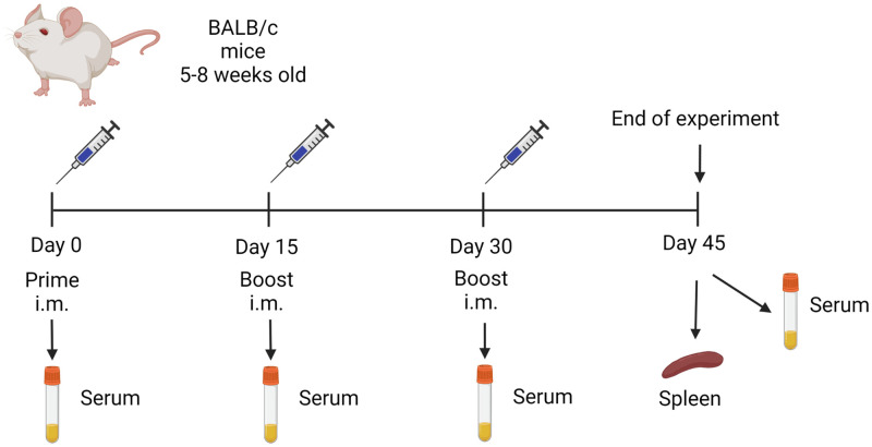
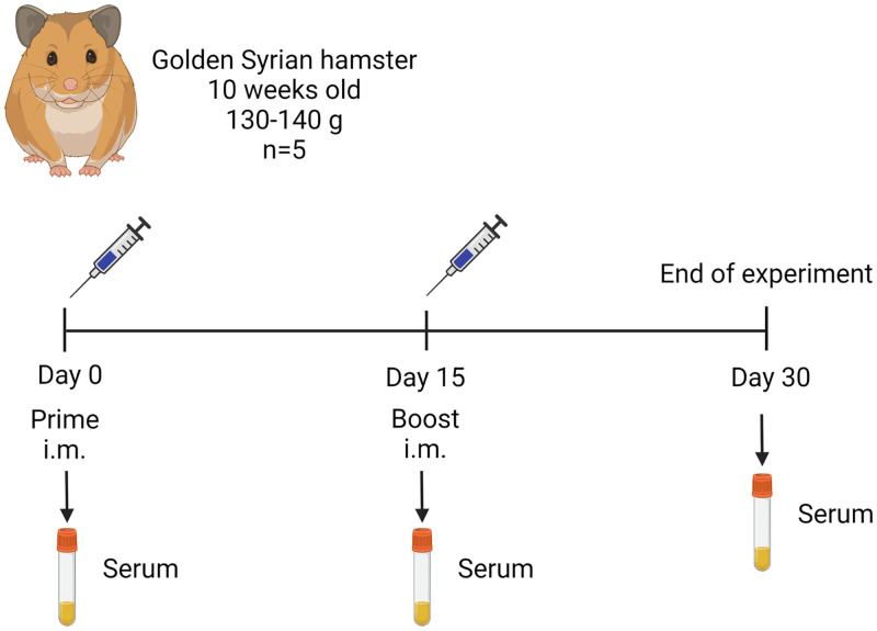
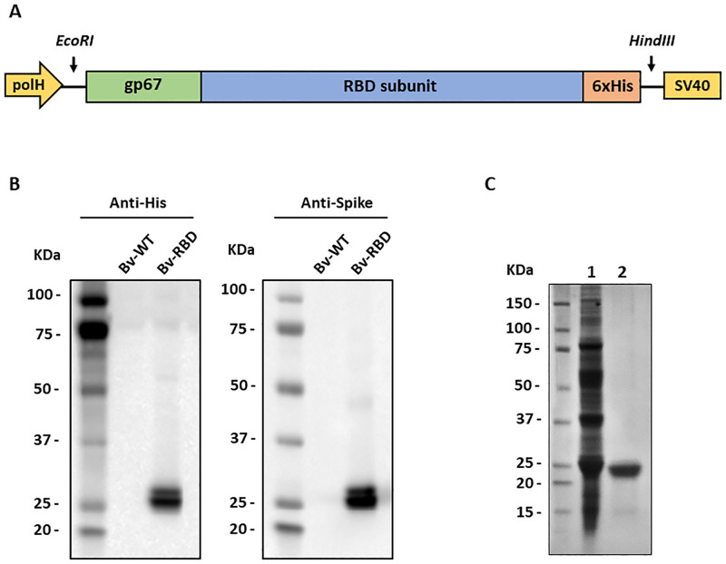
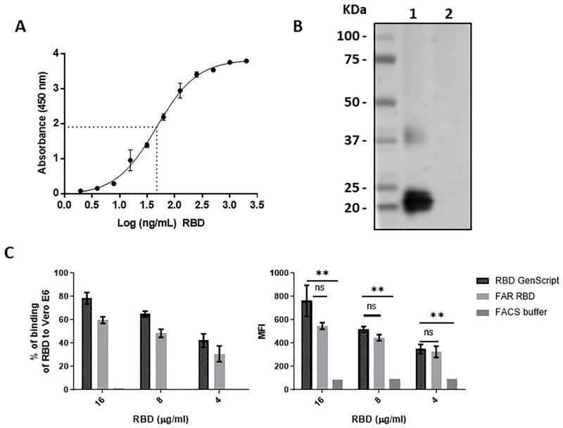
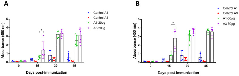
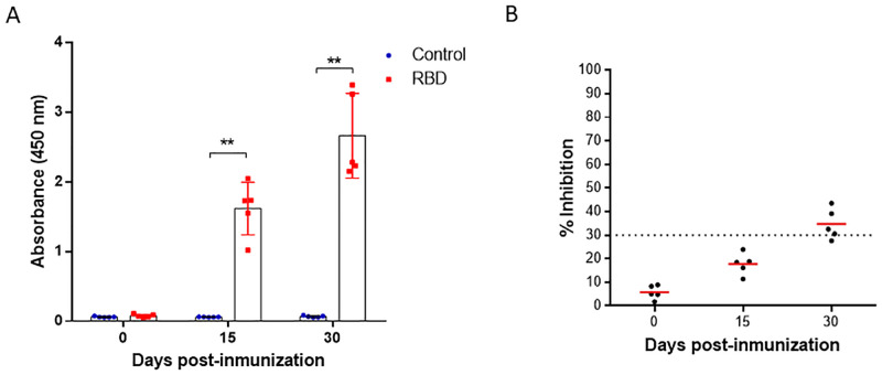
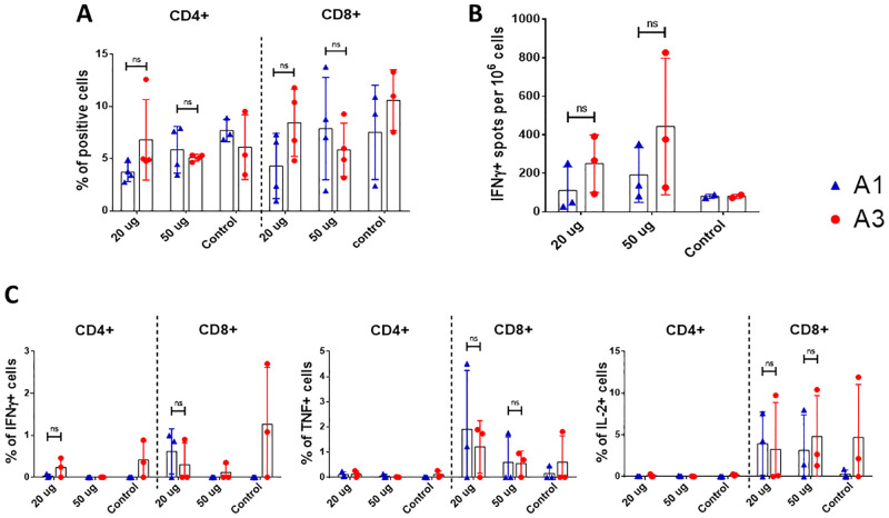
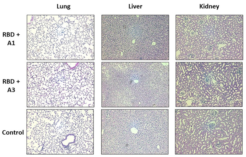
COVID-19 pandemic has accelerated the development of vaccines against its etiologic agent, SARS-CoV-2. However, the emergence of new variants of the virus lead to the generation of new alternatives to improve the current sub-unit vaccines in development. In the present report, the immunogenicity of the Spike RBD of SARS-CoV-2 formulated with an oil-in-water emulsion and a water-in-oil emulsion with squalene was evaluated in mice and hamsters. The RBD protein was expressed in insect cells and purified by chromatography until >95% purity. The protein was shown to have the appropriate folding as determined by ELISA and flow cytometry binding assays to its receptor, as well as by its detection by hamster immune anti-S1 sera under non-reducing conditions. In immunization assays, although the cellular immune response elicited by both adjuvants were similar, the formulation based in water-in-oil emulsion and squalene generated an earlier humoral response as determined by ELISA. Similarly, this formulation was able to stimulate neutralizing antibodies in hamsters. The vaccine candidate was shown to be safe, as demonstrated by the histopathological analysis in lungs, liver and kidney. These results have shown the potential of this formulation vaccine to be evaluated in a challenge against SARS-CoV-2 and determine its ability to confer protection.


Conflict of interest statement

The authors have declared that no competing interests exist.

Figures

Fig 1
Fig 1. Mice immunization flow chart.
Mice were immunized by the intramuscular route using a prime-boost regimen with a booster on days 15 and 30. Seven groups of mice were included: group 1 (20 μg RBD/A1, n = 5), group 2 (50 μg RBD/A1, n = 5), group 3 (20 μg RBD/A3, n = 5), group 4 (50 μg RBD/A3, n = 5), group 5 (only A1 n = 5), group 6 (only A3, n = 5) and group 7 (no immunization).
Fig 2
Fig 2. Hamster immunization flow chart.
Hamsters were immunized by the intramuscular route with 30 μg of purified RBD in adjuvant A3 using a prime-boost regimen with a booster on day 15.
Fig 3
Fig 3. RBD expression and purification.
(A) Design of the expression cassette integrated into the recombinant baculovirus. (B) Detection of RBD from infected culture supernatants using an anti-His (left) and anti-spike (right) antibody. Bv-WT: Wild type baculovirus; Bv-RBD: RBD expressing baculovirus. (C) SDS-PAGE of purified RBD after the affinity chromatography purification step (Lane 1) and size exclusion chromatography (Lane 2).
Fig 4
Fig 4. RBD binding and folding characterization in vitro.
(A) Dose dependent curve of RBD binding to human ACE-2 by ELISA, dashed lines represent the EC50 value. Dots and error bars represent the mean value of three independent experiments and the standard deviation, respectively. (B) Disulfide bond dependent recognition of RBD by hamsters immunized serum by Western blot. Lane1: RBD under non-reducing conditions; Lane 2: RBD under reducing conditions. (C) RBD binding to Vero E6 cell surface. The binding values are represented as the percentage of cells bound to RBD (left diagram) and the Mean Fluorescence Intensity (MFI) of each group was evaluated (right diagram). Two repetitions were performed per group, except in the FACS buffer group. Student t-test was used to compare the MFI values. ns: not significant (P>0.05); **: significant (P<0.01).
Fig 5
Fig 5. Detection of specific antibodies against RBD in mice.
Immunized mice were bled at 0, 15, 30 and 45 days post immunization. All sera were obtained by low-speed centrifugation. Serum samples were processed to detect specific antibodies against SARS-CoV-2 RBD protein using indirect ELISA assay. (A) Group immunized with 20 μg of RBD mixed with A1 and A3 (B) Group immunized with RBD 50 μg of RBD mixed with A1 and A4. Two-way ANOVA and post-hoc Tukey’s test were performed. *: P<0.01.
Fig 6
Fig 6. Detection of specific antibodies against RBD and neutralizing antibodies in hamsters.
(A) Immunized hamsters were bled at 0, 15 and 30 days post immunization. Serum samples were processed to detect specific antibodies against SARS-CoV-2 RBD protein using indirect ELISA assay. (B) Serum samples were processed to evaluate the neutralizing antibody titers against SARS-CoV-2 using sVNT. The cut-off for positive/negative neutralizing antibodies in the sample was 30% of inhibition of RBD binding to ACE-2. Two-way ANOVA and post-hoc Tukey’s test were performed. **: P<0.0001.
Fig 7
Fig 7. Evaluation of cellular immunity in mice vaccinated with purified RBD.
Mice were immunized with 20 and 50 μg of RBD using two different adjuvants (A1 and A3) at 0, 15 and 30 days post immunization. On day 45 post-immunization mice were euthanized and spleens were processed. (A) Percentage of CD4 and CD8 positive cells by flow cytometry, between the groups immunized (n = 3, except the adjuvant control). (B) IFN-γ ELISPOT of splenocytes between the groups immunized (n = 3, except the adjuvant control). (C) Intracellular staining of Th1 cytokines (IFN-γ, TNF-α and IL-2) of splenocytes stimulated with RBD (n = 3, except the adjuvant control). ns: not significant (P>0.05), *: P<0.05.
Fig 8
Fig 8. Histopathological analysis of mice inoculated with purified RBD and control.
Organs were obtained 45 days after the first immunization and stained with hematoxylin-eosin (H&E). These images are representative slides from vaccinated mice and negative control mice. (A) Lung sections. (B) Liver sections. (C) Kidney sections. All the images are in a 200X magnification.

References

    1. Zhou P, Yang X-L, Wang X-G, Hu B, Zhang L, Zhang W, et al.. A pneumonia outbreak associated with a new coronavirus of probable bat origin. Nature. 2020;579: 270–273. doi: 10.1038/s41586-020-2012-7 - DOI - PMC - PubMed
    1. Walls AC, Park Y-J, Tortorici MA, Wall A, McGuire AT, Veesler D. Structure, Function, and Antigenicity of the SARS-CoV-2 Spike Glycoprotein. Cell. 2020; 1–12. doi: 10.1016/j.cell.2020.02.058 - DOI - PMC - PubMed
    1. Lan J, Ge J, Yu J, Shan S, Zhou H, Fan S, et al.. Structure of the SARS-CoV-2 spike receptor-binding domain bound to the ACE2 receptor. Nat 2020 5817807. 2020;581: 215–220. doi: 10.1038/s41586-020-2180-5 - DOI - PubMed
    1. Tai W, He L, Zhang X, Pu J, Voronin D, Jiang S, et al.. Characterization of the receptor-binding domain (RBD) of 2019 novel coronavirus: implication for development of RBD protein as a viral attachment inhibitor and vaccine. Cell Mol Immunol 2020 176. 2020;17: 613–620. doi: 10.1038/s41423-020-0400-4 - DOI - PMC - PubMed
    1. Ou J, Zhou Z, Dai R, Zhang J, Zhao S, Wu X, et al.. V367F Mutation in SARS-CoV-2 Spike RBD Emerging during the Early Transmission Phase Enhances Viral Infectivity through Increased Human ACE2 Receptor Binding Affinity. J Virol. 2021;95. doi: 10.1128/JVI.00617-21 - DOI - PMC - PubMed

Publication types