. 2022 Sep 27;16(9):14210-14229.
doi: 10.1021/acsnano.2c04330. Epub 2022 Aug 23.

Barriers to the Intestinal Absorption of Four Insulin-Loaded Arginine-Rich Nanoparticles in Human and Rat

Affiliations

Barriers to the Intestinal Absorption of Four Insulin-Loaded Arginine-Rich Nanoparticles in Human and Rat

Patrik Lundquist et al. ACS Nano. .

Abstract

Peptide drugs and biologics provide opportunities for treatments of many diseases. However, due to their poor stability and permeability in the gastrointestinal tract, the oral bioavailability of peptide drugs is negligible. Nanoparticle formulations have been proposed to circumvent these hurdles, but systemic exposure of orally administered peptide drugs has remained elusive. In this study, we investigated the absorption mechanisms of four insulin-loaded arginine-rich nanoparticles displaying differing composition and surface characteristics, developed within the pan-European consortium TRANS-INT. The transport mechanisms and major barriers to nanoparticle permeability were investigated in freshly isolated human jejunal tissue. Cytokine release profiles and standard toxicity markers indicated that the nanoparticles were nontoxic. Three out of four nanoparticles displayed pronounced binding to the mucus layer and did not reach the epithelium. One nanoparticle composed of a mucus inert shell and cell-penetrating octarginine (ENCP), showed significant uptake by the intestinal epithelium corresponding to 28 ± 9% of the administered nanoparticle dose, as determined by super-resolution microscopy. Only a small fraction of nanoparticles taken up by epithelia went on to be transcytosed via a dynamin-dependent process. In situ studies in intact rat jejunal loops confirmed the results from human tissue regarding mucus binding, epithelial uptake, and negligible insulin bioavailability. In conclusion, while none of the four arginine-rich nanoparticles supported systemic insulin delivery, ENCP displayed a consistently high uptake along the intestinal villi. It is proposed that ENCP should be further investigated for local delivery of therapeutics to the intestinal mucosa.

Keywords: human; insulin; jejunum; nanoparticle; oral peptide delivery.


Conflict of interest statement

The authors declare no competing financial interest.

Figures

Figure 1
Figure 1
A. TRANS-INT NP evaluation criteria. NP were prioritized according to criteria of size (<350 nm diameter), negative or approximately neutral surface charge (ζ-potential), loading efficiency (incorporation of more than 50% of added peptide), final loading of peptide (>1% by weight), stability of formulation with regard to dissolution and stability in simulated intestinal fluids, relevant buffers, cell culture media, and controlled release of peptide cargos in these media., NP passing these criteria were evaluated in initial in vitro tests including protection of cargo against proteases, and toxicity in Caco-2 cell culture. After evaluation, four TRANS-INT NP were selected for further in vitro, ex vivo and in situ evaluation in this study. (B) Study workflow. Arginine-rich NP selected for inclusion in this study were resynthesized and evaluated according to TRANS-INT criteria. NP components were fluorescently labeled. NP permeability and interactions with Caco-2 cells were investigated. NP were characterized in isolated human jejunal tissues mounted in an Ussing chamber ex vivo, and in rat jejunal loops in situ. Permeability and tissue interactions of the NP were investigated, and PKPD of the insulin formulation was monitored in the rat circulation.
Figure 2
Figure 2
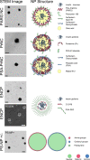
Schematics and micrographs of proposed NP structures. Electron micrographs of the included NP are shown. Sketches of NP structures are illustrations of NP composition and not exact models. The sketches depict a basic model of the NP core–shell structure indicating the localization of insulin and the assumed composition of the NP surface. Actual structures are likely to be more complex and dynamic. Components are not to scale. NP components are color-coded by charge. Fluid oil phases are indicated in yellow. Scale bars in black or white represent 200 nm. Abbreviations: STEM, scanning transmission electron microscopy; FESEM, field emission scanning electron microscopy.
Figure 3
Figure 3
NP–tissue interactions in human jejunum. Images depict the distribution of NP in human jejunal mucosa with a mucus layer in Ussing chambers at 90 min after NP administration to the mucosal side. In all images, the NPs are stained green, actin is stained red, cell membranes are stained purple, and nuclei are stained cyan. The scale bar depicts 50 μm. (A) PSA-PrNC (green), (B) PrNC (green). NPs in panels A and B show pronounced mucus binding and little interaction with the epithelium. (C) PrNC (green) and PSA-PrNC (not shown) were in rare cases found below the epithelial surface. At these sites, the epithelial cell layer was missing, and hence, the epithelial barrier was defective as indicated by the lack of actin staining of the apical BBM (red). The defect is indicated by a white bracket. One single villus is shown. Figure S7 shows another example of PrNC localized in the lamina propria below an epithelial defect. (D) ENCP (green) showed little to no interaction with the mucus but was found at the apical BBM of jejunal enterocytes, creating a green outline of the epithelial surface (see Figure 4 for higher resolution). (E) Actin staining outlines the brush-border membrane in red. (F) Nearly total colocalization of the ENCP signal (green) and brush-border membrane actin stain in red, resulting in an orange overlay indicating ENCP localization to the BBM. (G) PS-NP+ (green) and (H) PS-NP– (green); both polystyrene control NP, PS-NP+ and PS-NP–, showed pronounced mucus binding and little to no interaction with the epithelium. All NP were imaged in at least five sections in tissue specimens derived from at least two parallel Ussing chambers from each of two donors.
Figure 4
Figure 4
Localization of ENCP in enterocyte brush-border membranes. The LSM image shows that the vast majority of endocytosed ENCPs are localized in the enterocyte brush-border membrane after 90 min of exposure in the Ussing chamber. (A) Overlay of all channels. Nuclei stained with DAPI. (B) Membrane staining in purple by wheat germ agglutinin. (C) ENCPs in green are localized to the brush-border membranes (BBM). Some endocytosed nanoparticles are seen in punctate stain just beneath the epithelial BBM. (D) Red phalloidin staining of actin visualizing the BBM. ENCP was imaged in several sections in tissue specimens from two parallel Ussing chambers from two donors.
Figure 5
Figure 5
Quantitation of internalized ENCP in jejunal epithelial enterocytes. (A) SIM imaging allows quantitation of ENCPs in enterocytes. When added to the apical side of jejunum, ENCP were primarily found in the BBM with few particles in the basal regions of the cells and nearly no NPs in the subepithelium. All enterocytes internalized ENCPs, but no NPs were detected in goblet cells (black lacuna). ENCP labeled in green. (B) Approximately 75% of the villi surface had absorbed ENCP (green) after 90 min exposure. Actin staining was omitted for clarity. (C) Quantitation of the number of ENCP absorbed per unit area in villi in tissues from four donors. Values are given as average ± SD. (D) ENCP (green) were, after apical addition, localized to the BBM and were internalized into punctate stains reminiscent of early endosomes. (E) Free FITC-C12-R8 polymer (green) stained the BBM but did not enter endosomes. (F) When added to the basolateral chamber ENCP (green) diffused unhindered through the lamina propria to the epithelial tight junctions at the upper basolateral edge of enterocytes (arrows). No ENCP were visualized within the BBM. In all LSM images (B–F), ENCP or FITC-C12-R8 are stained green, actin is stained red, and nuclei are stained cyan; in addition, in panels D and E, cell membranes are stained purple. All sections were processed after 90 min in the Ussing chambers.
Figure 6
Figure 6
ENCP permeability mechanism across human jejunal mucosa. (A) Permeability of HRP in the presence and absence of the dynasore. (B) Dynasore reduced permeability of FD3. (C) Reduced endocytosis of FD3 in the presence of dynasore illustrated by confocal microscopy. FD3 in red; nuclei stained in cyan. (D, E) Dynasore effects on ENCP Papp and permeability. (F) Dynasore seems to reduce internalization of ENCPs into enterocytes. ENCP in green. Images from control and dynasore treated tissue specimens were imagined and processed using identical settings. Average ± SD is shown, n = 3, ** p < 0.01, *** p < 0.001.
Figure 7
Figure 7
In vivo administration of insulin-loaded NPs to rat jejunal loops. Pharmacodynamics of blood glucose responses: (A) PARG NC; (B) PrNC; and (C) ENCPs. (D) Concentrations of human insulin in blood in dosed rats. (E) Relative bioavailabilities for NP administered to rat jejunal loops compared to sc administration (Fsc = 100%). (F) Confocal microscopy of fluorescent NP interactions with the rat jejunal mucosa. PrNC in red. ENCP in green. Scale bar equals 50 μm. Average ± SEM is shown, NS = not significant.

Similar articles

Cited by

References

    1. Lundquist P.; Artursson P. Oral Absorption of Peptides and Nanoparticles across the Human Intestine: Opportunities, Limitations and Studies in Human Tissues. Adv. Drug Delivery Rev. 2016, 106, 256–276. 10.1016/j.addr.2016.07.007. - DOI - PubMed
    1. Brayden D. J.; Hill T.; Fairlie D.; Maher S.; Mrsny R. Systemic Delivery of Peptides by the Oral Route: Formulation and Medicinal Chemistry Approaches. Adv. Drug Delivery Rev. 2020, 157, 2–36. 10.1016/j.addr.2020.05.007. - DOI - PubMed
    1. Durán-Lobato M.; Niu Z.; Alonso M. J. Oral Delivery of Biologics for Precision Medicine. Adv. Mater. 2020, 32 (13), 1901935.10.1002/adma.201901935. - DOI - PubMed
    1. Brayden D. J. The Centenary of the Discovery of Insulin: An Update on the Quest for Oral Delivery. Front. Drug Delivery 2021, 1, 1.10.3389/fddev.2021.726675. - DOI
    1. Couvreur P. Polyalkylcyanoacrylates as Colloidal Drug Carriers. Crit. Rev. Ther. Drug Carrier Syst. 1988, 5 (1), 1–20. - PubMed

Publication types