. 2022 Oct 10;32(19):4286-4298.e5.
doi: 10.1016/j.cub.2022.07.064. Epub 2022 Aug 22.

The transcription factor Tbx5 regulates direction-selective retinal ganglion cell development and image stabilization

Affiliations

The transcription factor Tbx5 regulates direction-selective retinal ganglion cell development and image stabilization

Timour Al-Khindi et al. Curr Biol. .

Abstract

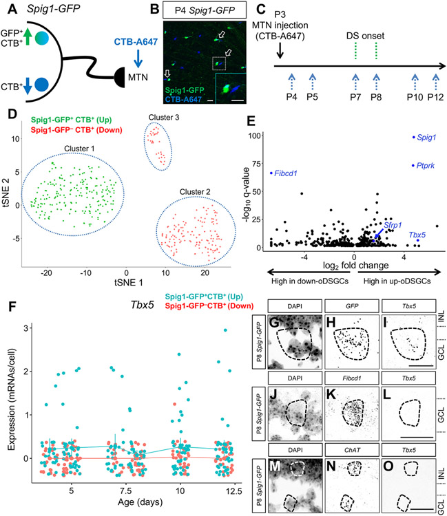
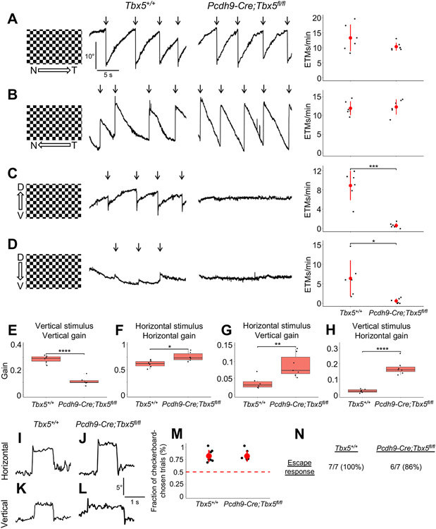
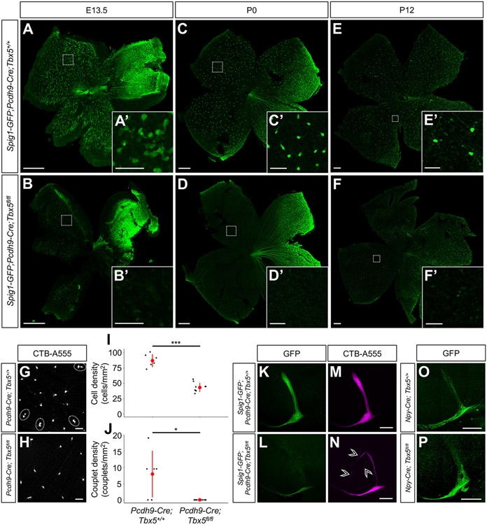
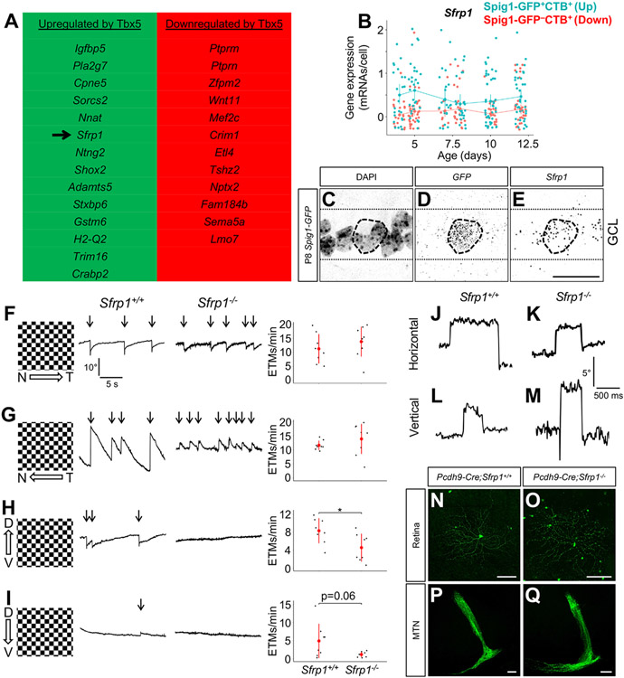
The diversity of visual input processed by the mammalian visual system requires the generation of many distinct retinal ganglion cell (RGC) types, each tuned to a particular feature. The molecular code needed to generate this cell-type diversity is poorly understood. Here, we focus on the molecules needed to specify one type of retinal cell: the upward-preferring ON direction-selective ganglion cell (up-oDSGC) of the mouse visual system. Single-cell transcriptomic profiling of up- and down-oDSGCs shows that the transcription factor Tbx5 is selectively expressed in up-oDSGCs. The loss of Tbx5 in up-oDSGCs results in a selective defect in the formation of up-oDSGCs and a corresponding inability to detect vertical motion. A downstream effector of Tbx5, Sfrp1, is also critical for vertical motion detection but not up-oDSGC formation. These results advance our understanding of the molecular mechanisms that specify a rare retinal cell type and show how disrupting this specification leads to a corresponding defect in neural circuitry and behavior.

Keywords: direction-selectivity; eye movements; neural development; retina; retinal ganglion cell; single-cell RNA-seq.


Conflict of interest statement

Declaration of interests The authors declare no competing interests.

Figures

Figure 1.
Figure 1.. Transcriptomic profiling of MTN-projecting ON DSGCs.
(A) Upward-preferring Spig1-GFP+ and downward-preferring Spig1-GFP ON direction-selective ganglion cells (oDSGCs) were retrogradely labeled by stereotactic injection of CTB-A647 into the medial terminal nucleus (MTN). (B) Whole-mount retina at P4 shows GFP+CTB+ up and GFPCTB+ down oDSGCs after successful MTN targeting at P3. Note the couplets of retrogradely labeled RGCS (arrows) that are differentially labeled (inset). (C) Experimental design for single-cell RNA sequencing. MTN injections were done at P3. Cells were collected before (P4, P5), during (P7, P8), and after (P10, P12) DS onset. (D) tSNE plot of single-cell RNA sequencing data reveals 3 RGC populations: 2 groups of MTN-projecting oDSGCs (Spig1-GFP+CTB+ cells–cluster 1 and Spig1-GFP CTB+ cells–cluster 2) and 1 group that likely corresponds to ipRGCs (Spig1-GFPCTB+ cells–cluster 3). (E) Volcano plot showing q-values and effect sizes for genes that are significantly differentially expressed between up- and down-oDSGCs (all genes shown have q<0.05). Spig1, Ptprk, Sfrp1, and Tbx5 were differentially expressed in up-oDSGCs, whereas Fibcd1 was differentially expressed in down-oDSGCs. (F) Tbx5 expression in up-oDSGCs was constant from P4 through P12 and was absent in GFP CTB+ down-oDSGCs. Note the extremely low Tbx5 transcript number per cell and the small fraction of Spig1-GFP+CTB+ RGCs that express Tbx5 at any given time point. Data presented as mean ± 95% confidence intervals. (G-O) RNAscope in situ hybridization in P8 Spig1-GFP retinas shows Tbx5 expression in GFP+ up-oDSGCs (G-I), absent Tbx5 expression in Fibcd1+ down-oDSGCs (J-L), and absent Tbx5 expression in ChAT+ SACs (M-O). GCL, ganglion cell layer. INL, inner nuclear layer. Scale bars, 25 um. See also Figure S1.
Figure 2.
Figure 2.. Pcdh9-Cre;Tbx5fl/fl mice show selective defects in vertical motion detection.
(A-D) Optokinetic reflex (OKR) results in adult Tbx5+/+ and Pcdh9-Cre;Tbx5fl/fl mice in response to nasotemporal (A), temporonasal (B), ventrodorsal (C), or dorsoventral (D) continuous motion, measured by scoring eye tracking movements (ETMs) per minute (arrows). Pcdh9-Cre;Tbx5fl/fl mice exhibited absent OKRs in both the ventrodorsal and dorsoventral directions. Data presented as mean ± SD. (E-H) Pcdh9-Cre;Tbx5fl/fl animals showed reduced gain in response to vertical sinusoidal stimuli (E) and enhanced gain in response to horizontal sinusoidal stimuli (F). Pcdh9-Cre;Tbx5fl/fl mice demonstrated enhanced vertical gain in response to horizontal stimuli (G) and enhanced horizontal gain in response to vertical stimuli (H). Data presented as mean ± IQR with whiskers representing the range. (I-L) Tbx5+/+ and Pcdh9-Cre;Tbx5fl/fl mice both performed voluntary horizontal and vertical saccades. (M) Pcdh9-Cre;Tbx5fl/fl mice showed normal performance on the visual cliff task, a measure of depth perception (dashed line represents chance performance). Data presented as mean ± SD. (N) Pcdh9-Cre;Tbx5fl/fl mice showed normal escape behaviors on the looming task, a measure of the ability to detect overhead looming stimuli. N, nasal. T, temporal. D, dorsal. V, ventral. *p < 0.05; **p < 0.01; ***p < 0.001; ****p < 0.0001. See also Figure S2.
Figure 3.
Figure 3.. Up-oDSGCs are absent in Spig1-GFP;Pcdh9-Cre;Tbx5fl/fl mice.
(A-F) Spig1-GFP+ up-oDSGCs were absent in Spig1-GFP;Pcdh9-Cre;Tbx5fl/fl mice at E13.5 (A and D), P0 (B and E), and P12 (C and F). (G-H) Whole-mount retinas of P6 Pcdh9-Cre;Tbx5+/+ (G) and Pcdh9-Cre;Tbx5fl/fl (H) retinas are shown following retrograde injection of CTB-555 into the MTN at P4. RGC couplets, corresponding to 1 up- and 1 down-oDSGC, were observed in Pcdh9-Cre;Tbx5+/+ retinas (dashed circles in G), but were absent in Pcdh9-Cre;Tbx5fl/fl retinas. (I-J) P6 Pcdh9-Cre;Tbx5fl/fl mice showed a ~50% reduction in MTN-projecting RGC density (I) and absence of up- and down-oDSGC couplets (J). Data presented as mean ± SD. (K-N) Following intraocular CTB-A555 injections at P10, the dorsal MTN was hypoinnervated in P12 Spig1-GFP;Pcdh9-Cre;Tbx5fl/fl mice, while the ventral MTN was normally innervated. Mistargeted CTB+ axons were observed in the proximity of the MTN in Spig1-GFP;Pcdh9-Cre;Tbx5fl/fl mice (arrowheads in N). (O-P) The MTN was appropriately innervated in adult Npy-Cre;Tbx5fl/fl mice following intraocular AAV2 FLEX-GFP injection. Scale bars, 250 um in (A-F) and (K-P); 50 um in (A’-F’) and (G-H). *p < 0.05; ***p < 0.001. See also Figure S3.
Figure 4.
Figure 4.. Sfrp1 is a potential downstream effector of Tbx5.
(A) Cross-referencing bulk RNAseq data from Tbx5+/+ and Tbx5−/− cardiac tissue with our oDSGC single-cell RNAseq data yielded a list of genes potentially upregulated or downregulated by Tbx5 (rank-ordered by q-value). (B) Sfrp1 was expressed at slightly higher levels in up-oDSGCs compared to down-oDSGCs between P4 and P12. Data presented as mean ± 95% confidence intervals. (C-E) RNAscope in situ hybridization in P8 Spig1-GFP retinas shows Sfrp1 expression in GFP+ up-oDSGCs within the ganglion cell layer (GCL). (F-I) Optokinetic reflex (OKR) measurements in adult Sfrp1+/+ and Sfrpt−/− mice showed comparable performance in response to nasotemporal (F) and temporonasal (G) motion. Sfrpt−/− mice showed impaired OKR performance in response to ventrodorsal motion (H) and a trend to statistically significant impairment in response to dorsoventral motion (I). Data presented as mean ± SD. (J-M) Voluntary horizontal (J-K) and vertical (L-M) saccades were intact in Sfrp1−/− mice. (N-Q) Adult up-oDSGCs, labeled with intraocular AAV2 FLEX-GFP in Pcdh9-Cre;Sfrp1+/+ and Pcdh9-Cre;Sfrp1−/− mice, appeared grossly normal with regards to dendrite morphology (N-O) and axon projections to the MTN (P-Q). Scale bars, 25 um in (C-E); 100 um in (N-Q). *p < 0.05. ETM, eye-tracking movement. See also Figure S4.

References

    1. Rheaume BA, Jereen A, Bolisetty M, Sajid MS, Yang Y, Renna K, Sun L, Robson P, and Trakhtenberg EF (2018). Single cell transcriptome profiling of retinal ganglion cells identifies cellular subtypes. Nat. Commun 9, 2759. 10.1038/S41467-018-05134-3. - DOI - PMC - PubMed
    1. Tran NM, Shekhar K, Whitney IE, Jacobi A, Benhar I, Hong G, Yan W, Adiconis X, Arnold ME, Lee JM, Levin JZ, Lin D, Wang C, Lieber CM, Regev A, He Z, and Sanes JR (2019). Single-cell profiles of retinal ganglion cells differing in resilience to injury reveal neuroprotective genes. Neuron 104, 1039–1055.e12. 10.1016/j.neuron.2019.11.006. - DOI - PMC - PubMed
    1. Wang F, Li E, De L, Wu Q, and Zhang Y (2021). OFF-transient alpha RGCs mediate looming triggered innate defensive response. Curr. Biol 31, 2263–2273.e3. 10.1016/j.cub.2021.03.025. - DOI - PubMed
    1. Nguyen-Ba-Charvet KT, and Rebsam A (2020). Neurogenesis and specification of retinal ganglion cells. Int. J. Mol. Sci 21, 451. 10.3390/ijms21020451. - DOI - PMC - PubMed
    1. Brodie-Kommit J, Clark BS, Shi Q, Shiau F, Kim DW, Langel J, Sheely C, Ruzycki PA, Fries M, Javed A, Cayouette M, Schmidt T, Badea T, Glaser T, Zhao H, Singer J, Blackshaw S, and Hattar S (2021). Atoh7-independent specification of retinal ganglion cell identity. Sci. Adv 7, eabe4983. 10.1126/sciadv.abe4983. - DOI - PMC - PubMed

Publication types

Substances